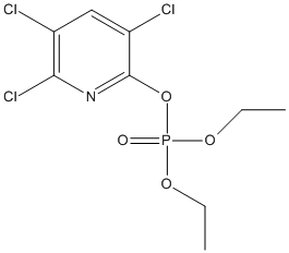
8 moreTitle: Chlorpyrifos oxon promotes tubulin aggregation via isopeptide cross-linking between diethoxyphospho-Lys and Glu or Asp: Implications for neurotoxicity Schopfer LM, Lockridge O Ref: Journal of Biological Chemistry, 293:13566, 2018 : PubMed
Exposure to organophosphorus toxicants (OP) can have chronic adverse effects that are not explained by inhibition of acetylcholinesterase, the cause of acute OP toxicity. We therefore hypothesized that OP-induced chronic illness is initiated by the formation of organophosphorus adducts on lysine residues in proteins, followed by protein cross-linking and aggregation. Here, Western blots revealed that exposure to the OP chlorpyrifos oxon converted porcine tubulin from its original 55-kDa mass to high-molecular-weight aggregates. Liquid chromatography-tandem MS analysis of trypsin-digested samples identified several diethoxyphospho-lysine residues in the OP-treated tubulin. Using a search approach based on the Batch Tag program, we identified cross-linked peptides and found that these chemically activated lysines reacted with acidic amino acid residues creating gamma-glutamyl--lysine or aspartyl--lysine isopeptide bonds between beta- and alpha-tubulin. Of note, these cross-linked tubulin molecules accounted for the high-molecular-weight aggregates. To the best of our knowledge, this is the first report indicating that chlorpyrifos oxon-exposed tubulin protein forms intermolecular cross-links with other tubulin molecules, resulting in high-molecular-weight protein aggregates. It is tempting to speculate that chronic illness from OP exposure may be explained by a mechanism that starts with OP adduct formation on protein lysines followed by protein cross-linking. We further speculate that OP-modified or cross-linked tubulin can impair axonal transport, reduce neuron connections, and result in neurotoxicity.
        
Title: Mice treated with chlorpyrifos or chlorpyrifos oxon have organophosphorylated tubulin in the brain and disrupted microtubule structures, suggesting a role for tubulin in neurotoxicity associated with exposure to organophosphorus agents Jiang W, Duysen EG, Hansen H, Shlyakhtenko L, Schopfer LM, Lockridge O Ref: Toxicol Sci, 115:183, 2010 : PubMed
Exposure to organophosphorus (OP) agents can lead to learning and memory deficits. Disruption of axonal transport has been proposed as a possible explanation. Microtubules are an essential component of axonal transport. In vitro studies have demonstrated that OP agents react with tubulin and disrupt the structure of microtubules. Our goal was to determine whether in vivo exposure affects microtubule structure. One group of mice was treated daily for 14 days with a dose of chlorpyrifos that did not significantly inhibit acetylcholinesterase. Beta-tubulin from the brains of these mice was diethoxyphosphorylated on tyrosine 281 in peptide GSQQY(281)RALTVPELTQQMFDSK. A second group of mice was treated with a single sublethal dose of chlorpyrifos oxon (CPO). Microtubules and cosedimenting proteins from the brains of these mice were visualized by atomic force microscopy nanoimaging and by Coomassie blue staining of polyacrylamide gel electrophoresis bands. Proteins in gel slices were identified by mass spectrometry. Nanoimaging showed that microtubules from control mice were decorated with many proteins, whereas microtubules from CPO-treated mice had fewer associated proteins, a result confirmed by mass spectrometry of proteins extracted from gel slices. The dimensions of microtubules from CPO-treated mice (height 8.7 +/- 3.1 nm and width 36.5 +/- 15.5 nm) were about 60% of those from control mice (height 13.6 +/- 3.6 nm and width 64.8 +/- 15.9 nm). A third group of mice was treated with six sublethal doses of CPO over 50.15 h. Mass spectrometry identified diethoxyphosphorylated serine 338 in peptide NS(338)NFVEWIPNNVK of beta-tubulin. In conclusion, microtubules from mice exposed to chlorpyrifos or to CPO have covalently modified amino acids and abnormal structure, suggesting disruption of microtubule function. Covalent binding of CPO to tubulin and to tubulin-associated proteins is a potential mechanism of neurotoxicity.
        
Title: Degradation by rat tissues in vitro of organophosphorus esters which inhibit cholinesterase Pla A, Johnson MK Ref: Biochemical Pharmacology, 38:1527, 1989 : PubMed
Hydrolytic "A"-esterase activities of various tissues of rat (plasma, liver, kidney, brain and intestinal mucosa) against selected OP esters of diverse structure as potential substrates (paraoxon, di-n-propyl paraoxon, di-n-butyl paraoxon, chlorpyrifos oxon, di-(4-phenyl butyl) phosphorofluoridate and the chiral isomers of ethyl 4-nitrophenyl phenylphosphonate) were studied. We have developed a sensitive and widely applicable assay depending on measuring decline in residual inhibitory power of any chosen OP against horse serum cholinesterase: for seven compounds examined so far I50s against BCHE ranged from 0.07 to 70 nM, and it is easy to monitor loss of OP starting from an initial 25 microM concentration. Progressive destruction rates were always highest in liver and plasma with activity sometimes detectable in kidney, brain but not in intestinal mucosa, but the ratios of activity between tissues differed for different substrates. At 25 microM/37 degrees/pH 7.2 hydrolysis rates ranged from 8500 nmol/min/g liver for di-(4-phenylbutyl) phosphorofluoridate down to 0.8 nmol/min for the butyl analogue of paraoxon; the rate for L(-) isomer of EPN oxon (23 nmol/min/g liver) was greater than 2x that for the D(+) isomer and for paraoxon. From our data we conclude that several OP hydrolases exist whose identity may be further characterised by use of selective substrates
        
8 lessTitle: Chlorpyrifos oxon promotes tubulin aggregation via isopeptide cross-linking between diethoxyphospho-Lys and Glu or Asp: Implications for neurotoxicity Schopfer LM, Lockridge O Ref: Journal of Biological Chemistry, 293:13566, 2018 : PubMed
Exposure to organophosphorus toxicants (OP) can have chronic adverse effects that are not explained by inhibition of acetylcholinesterase, the cause of acute OP toxicity. We therefore hypothesized that OP-induced chronic illness is initiated by the formation of organophosphorus adducts on lysine residues in proteins, followed by protein cross-linking and aggregation. Here, Western blots revealed that exposure to the OP chlorpyrifos oxon converted porcine tubulin from its original 55-kDa mass to high-molecular-weight aggregates. Liquid chromatography-tandem MS analysis of trypsin-digested samples identified several diethoxyphospho-lysine residues in the OP-treated tubulin. Using a search approach based on the Batch Tag program, we identified cross-linked peptides and found that these chemically activated lysines reacted with acidic amino acid residues creating gamma-glutamyl--lysine or aspartyl--lysine isopeptide bonds between beta- and alpha-tubulin. Of note, these cross-linked tubulin molecules accounted for the high-molecular-weight aggregates. To the best of our knowledge, this is the first report indicating that chlorpyrifos oxon-exposed tubulin protein forms intermolecular cross-links with other tubulin molecules, resulting in high-molecular-weight protein aggregates. It is tempting to speculate that chronic illness from OP exposure may be explained by a mechanism that starts with OP adduct formation on protein lysines followed by protein cross-linking. We further speculate that OP-modified or cross-linked tubulin can impair axonal transport, reduce neuron connections, and result in neurotoxicity.
        
Title: Mice treated with chlorpyrifos or chlorpyrifos oxon have organophosphorylated tubulin in the brain and disrupted microtubule structures, suggesting a role for tubulin in neurotoxicity associated with exposure to organophosphorus agents Jiang W, Duysen EG, Hansen H, Shlyakhtenko L, Schopfer LM, Lockridge O Ref: Toxicol Sci, 115:183, 2010 : PubMed
Exposure to organophosphorus (OP) agents can lead to learning and memory deficits. Disruption of axonal transport has been proposed as a possible explanation. Microtubules are an essential component of axonal transport. In vitro studies have demonstrated that OP agents react with tubulin and disrupt the structure of microtubules. Our goal was to determine whether in vivo exposure affects microtubule structure. One group of mice was treated daily for 14 days with a dose of chlorpyrifos that did not significantly inhibit acetylcholinesterase. Beta-tubulin from the brains of these mice was diethoxyphosphorylated on tyrosine 281 in peptide GSQQY(281)RALTVPELTQQMFDSK. A second group of mice was treated with a single sublethal dose of chlorpyrifos oxon (CPO). Microtubules and cosedimenting proteins from the brains of these mice were visualized by atomic force microscopy nanoimaging and by Coomassie blue staining of polyacrylamide gel electrophoresis bands. Proteins in gel slices were identified by mass spectrometry. Nanoimaging showed that microtubules from control mice were decorated with many proteins, whereas microtubules from CPO-treated mice had fewer associated proteins, a result confirmed by mass spectrometry of proteins extracted from gel slices. The dimensions of microtubules from CPO-treated mice (height 8.7 +/- 3.1 nm and width 36.5 +/- 15.5 nm) were about 60% of those from control mice (height 13.6 +/- 3.6 nm and width 64.8 +/- 15.9 nm). A third group of mice was treated with six sublethal doses of CPO over 50.15 h. Mass spectrometry identified diethoxyphosphorylated serine 338 in peptide NS(338)NFVEWIPNNVK of beta-tubulin. In conclusion, microtubules from mice exposed to chlorpyrifos or to CPO have covalently modified amino acids and abnormal structure, suggesting disruption of microtubule function. Covalent binding of CPO to tubulin and to tubulin-associated proteins is a potential mechanism of neurotoxicity.
        
Title: Blood acylpeptide hydrolase activity is a sensitive marker for exposure to some organophosphate toxicants Quistad GB, Klintenberg R, Casida JE Ref: Toxicol Sci, 86:291, 2005 : PubMed
Acylpeptide hydrolase (APH) unblocks N-acetyl peptides. It is a major serine hydrolase in rat blood, brain, and liver detected by derivatization with (3)H-diisopropyl fluorophosphate (DFP) or a biotinylated fluorophosphonate. Although APH does not appear to be a primary target of acute poisoning by organophosphorus (OP) compounds, the inhibitor specificity of this secondary target is largely unknown. This study fills the gap and emphasizes blood APH as a potential marker of OP exposure. The most potent in vitro inhibitors for human erythrocyte and mouse brain APH are DFP (IC(50) 11-17 nM), chlorpyrifos oxon (IC(50) 21-71 nM), dichlorvos (IC(50) 230-560 nM), naled (IC(50) 370-870 nM), and their analogs with modified alkyl substituents. (3)H-diisopropyl fluorophosphate is a potent inhibitor of mouse blood and brain APH in vivo (ED(50) 0.09-0.2 mg/kg and 0.02-0.03 mg/l for ip and vapor exposure, respectively). Mouse blood and brain APH and blood butyrylcholinesterase (BChE) are of similar sensitivity to DFP in vitro and in vivo (ip and vapor exposure), but APH inhibition is much more persistent in vivo (still >80% inhibition after 4 days). The inhibitory potency of OP pesticides in vivo in mice varies from APH selective (dichlorvos, naled, and trichlorfon), to APH and BChE selective (profenofos and tribufos), to ChE selective or nonselective (many commercial insecticides). Sarin administered ip at a lethal dose to guinea pigs inhibits blood acetylcholinesterase and BChE completely but erythrocyte APH only partially. Blood APH activity is therefore a sensitive marker for exposure to some but not all OP pesticides and chemical warfare agents.
        
Title: Inhibition of carboxylesterases in SH-SY5Y human and NB41A3 mouse neuroblastoma cells by organophosphorus esters Ehrich M, Correll L Ref: J Toxicol Environ Health, 53:385, 1998 : PubMed
Carboxylesterases (CbxE) can be inhibited by organophosphorus esters (OPs) without causing clinical evidence of toxicity. CbxE are thought to protect the critical enzyme acetylcholinesterase (AChE) from OP inhibition in animals. CbxE and AChE are both present in neuroblastoma cells, but, even though these cells have potential to be an in vitro model of OP toxicity, the effect of OPs on CbxE and the relationship of CbxE inhibition and AChE inhibition have not yet been examined in these cells. Therefore, this study examined concentration-related OP-induced inhibition of CbxE in human SH-SY5Y and mouse NB41A3 neuroblastoma cells with 11 active esterase inhibitors: paraoxon, malaoxon, chlorpyrifos-oxon, tolyl saligenin phosphate (TSP), phenyl saligenin phosphate (PSP), diisopropyl phosphorofluoridate (DFP), mipafox, dichlorvos, trichlorfon, dibutyryl dichlorovinyl phosphate (DBVP), and dioctyl dichlorovinyl phosphate (DOVP). All could inhibit CbxE, although the enzyme was less likely to be inhibited than AChE following exposure to 9 of the test compounds in the human cell line and to all 11 of the test compounds in the murine cell line. Species differences in concentration-related inhibitions of CbxE were evident. When cells were exposed first to an OP with a low IC50 toward CbxE (PSP), followed by an OP with high affinity for AChE (paraoxon or malaoxon), inhibitions of CbxE and AChE were additive. This indicated that CbxE did not protect AChE from OP-induced inhibition in this cell culture model.
        
Title: Rat brain acetylcholinesterase activity: developmental profile and maturational sensitivity to carbamate and organophosphorus inhibitors Mortensen SR, Hooper MJ, Padilla S Ref: Toxicology, 125:13, 1998 : PubMed
A growing body of evidence indicates that young animals exhibit an increased susceptibility to the lethal effects of cholinesterase (ChE)-inhibiting insecticides. Our laboratory is engaged in defining factors which may explain this age-related sensitivity. This report includes results from experiments designed to compare the developmental profiles, kinetic parameters and intrinsic (i.e. in vitro) sensitivity of developing male rat brain acetylcholinesterase (AChE) activity to carbamate and organophosphorus anticholinesterases. Total ChE activity in whole brain for each age was composed of about 90% AChE and 10% butyrylcholinesterase (BCHE) activity for the six ages examined. Brain AChE activity showed an age-related increase in Vmax until postnatal day 17 with no change in Km (average of all six ages approximately equal to 72 microM). Optimal substrate (acetylthiocholine) concentration for each age was 1 mM, and there was substrate inhibition (approximately 10%) at 2.5 mM. IC50s (the concentration of compound that inhibits 50% of the AChE activity in 30 min at 26 degrees C) defined concomitantly for postnatal day 4 and adult brain AChE using either aldicarb, carbaryl, chlorpyrifos-oxon or malaoxon were virtually identical at both ages with average IC50 values being: aldicarb = 2.4 microM, carbaryl = 1.7 microM, chlorpyrifos-oxon = 4.9 nM and malaoxon = 140 nM. In summary, AChE in young and adult brain differs mostly in specific activity while the Km(s), substrate profiles, and in vitro sensitivity to selected anticholinesterase insecticides are not different. Therefore, these data support the hypothesis that the greater sensitivity of the young animals to anticholinesterase pesticides is not due to the greater sensitivity of the target molecule AChE to these inhibitors.
        
Title: Kinetic analysis of the in vitro inhibition, aging, and reactivation of brain acetylcholinesterase from rat and channel catfish by paraoxon and chlorpyrifos-oxon Carr RL, Chambers JE Ref: Toxicology & Applied Pharmacology, 139:365, 1996 : PubMed
In rats, the phosphorothionate insecticide parathion exhibits greater toxicity than chlorpyrifos, while in catfish the toxicities are reversed. The in vitro inhibition of brain acetylcholinesterase (AChE) by the active metabolites of the insecticides and the rates at which these inhibitor-enzyme complexes undergo reactivation/ aging were investigated in both species. Rat AChE was more sensitive to inhibition than catfish AChE as demonstrated by greater bimolecular rate constants (ki) in rats than in catfish. In both species, chlorpyrifos-oxon yielded higher ki's than paraoxon. The higher association constant (KA) of chlorpyrifos-oxon than paraoxon in both species and the lack of significant differences in the phosphorylation constants (kp) suggest that association of the inhibitor with AChE is the principal factor in the different potencies between these two inhibitors. In catfish, the ki of chlorpyrifos-oxon was 22-fold greater than that of paraoxon, while in rats it was 9-fold greater, suggesting that target site sensitivity is an important factor in the higher toxicity of chlorpyrifos to catfish but not in the higher toxicity of parathion to rats. No spontaneous reactivation of phosphorylated catfish AChE occurred and there were no differences in the first oder aging constants (ka) between compounds. For phosphorylated rat AChE, there were no differences in the first order reactivation constants (kr) but the ka for chlorpyrifos-oxon was significantly greater than that for paraoxon. This difference suggests that the steric positioning of the diethyl phosphate in the esteratic site is not the same between the two compounds, leading to differences in aging.
        
Title: Identification and isolation of two rat serum proteins with A-esterase activity toward paraoxon and chlorpyrifos-oxon Pond AL, Coyne CP, Chambers HW, Chambers JE Ref: Biochemical Pharmacology, 52:363, 1996 : PubMed
The active metabolites (oxons) of phosphorothionate insecticides can be detoxified via A-esterase hydrolysis. Two enzymes with A-esterase activity have been isolated from rat serum. Whole serum was applied to anion exchange gel (DEAE Sepharose Fast Flow) and incubated (1 hr). Tris-HCl buffer (0.05 M; pH 7.7, at 5 degrees) containing 0.25 M NaCl was added to the slurry and incubated. The decant, containing low A-esterase activity but a high protein concentration, was discarded. Further displacement of A-esterase from DEAE gel was achieved with 1.0 M NaCl in 0.05 M Tris-HCl buffer (Ph 7.7 at 5 degrees). Following desalting and concentration, further separation was achieved by gel filtration (Sephacryl S-100 HR) and two sequential preparative scale isoelectric focusings. Final fractions contained two proteins of high molecular mass (one about 200 kDa and one between 137 and 200 kDa). The apparent range of isoelectric points for the two enzymes was 4.5 to 5.6. Following native-PAGE analysis, activity stains with beta-naphthyl acetate and Fast Garnet GBC in the presence of paraoxon (10-5 M) verified that A-esterase activity was associated with both proteins. Spectropho-tometric assay detected A-esterase activity toward paraoxon, chlorpyrifos-oxon, and phenyl acetate in the final preparation.
        
Title: Inhibition and aging of channel catfish brain acetylcholinesterase following exposure to two phosphorothionate insecticides and their active metabolites Carr RL, Straus DL, Chambers JE Ref: Journal of Toxicology & Environmental Health, 45:325, 1995 : PubMed
The inhibition and aging of acetylcholinesterase (AChE) in fingerling channel catfish (lctalurus punctatus) brain tissue was studied after single in vivo exposures to high levels of chlorpyrifos (0.25 mg/L), chlorpyrifos-oxon (7 micrograms/L), parathion (2.5 mg/L), or paraoxon (30 micrograms/L). Exposure to both parent compounds produced identical initial inhibition (95%), but in the later sampling times there was significantly more inhibited AChE in the chlorpyrifos-treated fish than in the parathion-treated fish (47% and 28%, respectively, on d 16). There were higher levels of aged AChE following chlorpyrifos exposure than following parathion exposure, but differences were not significant. Exposure to both oxons produced initial inhibition greater than 90%, and patterns of recovery and aging were statistically similar between both compounds; no significant inhibition was observed after d 11. The similar patterns of inhibition, recovery, and aging between the two oxon treatments, which have similar lipophilicities, suggest that the greater amount of AChE inhibition and aging observed in the chlorpyrifos-treated fish compared with the parathion-treated fish probably results from the higher lipophilicity of chlorpyrifos than of parathion. Overall, the prolonged brain AChE inhibition exhibited in catfish exposed to phosphorothionates is not the result of aging of the inhibited enzyme but is the result of either a slow rate or a lack of spontaneous reactivation.
        
Title: Inhibition patterns of brain acetylcholinesterase and hepatic and plasma aliesterases following exposures to three phosphorothionate insecticides and their oxons in rats Chambers JE, Carr RL Ref: Fundamental & Applied Toxicology, 21:111, 1993 : PubMed
Rats were administered high sublethal intraperitoneal dosages of the phosphorothionate insecticides parathion, methyl parathion, and chlorpyrifos, and their oxons. Acetylcholinesterase activities in cerebral cortex and medulla oblongata and aliesterase activities in liver and plasma were monitored at 2 hr and 1, 2, and 4 days after exposure. The maximal inhibition of brain acetylcholinesterase activity was not immediate with parathion and chlorpyrifos, reflecting the time required for bioactivation of the phosphorothionates as well as the effectiveness of the aliesterases to inactivate much of the hepatically generated oxons. In contrast, brain acetylcholinesterase activities were more quickly inhibited following administration of paraoxon and chlorpyrifos-oxon, which do not require bioactivation. Brain acetylcholinesterase was also rapidly inhibited following administration of methyl parathion and methyl paraoxon, reflecting the low sensitivity of the aliesterases to methyl paraoxon. Aliesterases were inhibited to a greater extent than acetylcholinesterase at each sampling time with parathion and chlorpyrifos and their oxons, whereas the reverse was true with methyl parathion and methyl paraoxon. All of the above patterns correlate with the in vitro sensitivities of acetylcholinesterase and aliesterases to the oxons. The very prolonged inhibition of esterase activities following chlorpyrifos treatment probably results from its substantially greater lipophilicity compared to the other compounds, which would allow it to be stored and released for gradual bioactivation. The data reported indicate that the disposition and effects of different phosphorothionate insecticides will be influenced by the sensitivities of target and nontarget esterases for their oxons and by their lipophilicity, and that predictions of in vivo responses can be made from in vitro data.
        
Title: Serum paraoxonase status: a major factor in determining resistance to organophosphates Li WF, Costa LG, Furlong CE Ref: Journal of Toxicology & Environmental Health, 40:337, 1993 : PubMed
A number of lines of evidence suggest that serum paraoxonase is protective against poisoning by organophosphorus substrates of this enzyme. Birds that have very low levels of paraoxon hydrolyzing activity in their sera are very susceptible to parathion poisoning. Rabbits, which have a sevenfold higher enzyme level compared with rats, have a fourfold higher resistance to paraoxon poisoning than rats. Rabbit paraoxonase hydrolyzes chlorpyrifos-oxon with a much higher turnover number than does rat paraoxonase, resulting in a very high resistance of rabbits to chlorpyrifos toxicity. Direct tests of paraoxonase protection have been carried out by injecting purified rabbit enzyme into rats. The protection achieved was higher for chlorpyrifos-oxon than for paraoxon, probably due to the high hydrolytic activity of the rabbit enzyme for chlorpyrifos-oxon. In humans, a substrate-dependent polymorphism of serum paraoxonase is observed, where one isoform of paraoxonase has a high turnover number for paraoxon and the other a low turnover number. Both isoforms appear to hydrolyze chlorpyrifos-oxon and phenylacetate at the same rate. Cloning and sequencing of the human paraoxonase cDNAs has elucidated the molecular basis of the polymorphism. Arginine at position 192 determines high paraoxonase activity, and glutamine at this position, low paraoxonase activity. In addition to the polymorphism, a 13-fold variation in serum enzyme levels within a given genetic class is seen. The experiments reported here demonstrate that rabbit paraoxonase injected into mice provides protection against the parent insecticide chlorpyrifos as well as the toxic oxon. These results suggest that serum paraoxonase status may serve as a biomarker for insecticide susceptibility in humans.
        
Title: Degradation by rat tissues in vitro of organophosphorus esters which inhibit cholinesterase Pla A, Johnson MK Ref: Biochemical Pharmacology, 38:1527, 1989 : PubMed
Hydrolytic "A"-esterase activities of various tissues of rat (plasma, liver, kidney, brain and intestinal mucosa) against selected OP esters of diverse structure as potential substrates (paraoxon, di-n-propyl paraoxon, di-n-butyl paraoxon, chlorpyrifos oxon, di-(4-phenyl butyl) phosphorofluoridate and the chiral isomers of ethyl 4-nitrophenyl phenylphosphonate) were studied. We have developed a sensitive and widely applicable assay depending on measuring decline in residual inhibitory power of any chosen OP against horse serum cholinesterase: for seven compounds examined so far I50s against BCHE ranged from 0.07 to 70 nM, and it is easy to monitor loss of OP starting from an initial 25 microM concentration. Progressive destruction rates were always highest in liver and plasma with activity sometimes detectable in kidney, brain but not in intestinal mucosa, but the ratios of activity between tissues differed for different substrates. At 25 microM/37 degrees/pH 7.2 hydrolysis rates ranged from 8500 nmol/min/g liver for di-(4-phenylbutyl) phosphorofluoridate down to 0.8 nmol/min for the butyl analogue of paraoxon; the rate for L(-) isomer of EPN oxon (23 nmol/min/g liver) was greater than 2x that for the D(+) isomer and for paraoxon. From our data we conclude that several OP hydrolases exist whose identity may be further characterised by use of selective substrates