Gallate
Tannins are natural polyphenolic compounds present in vascular plants. Hydrolyzable tannins are derivatives of gallic acid (3,4,5-trihydroxybenzoic acid) or its meta-depsidic forms that are esterified to polyol, catechin, or triterpenoid units. Gallate is product of hydrolysis of Ethyl-gallate. used in the pharmaceutical industry and is an important intermediate in the synthesis of the antibacterial agent trimethoprim
General
Type : Derivative of Gallate,Benzoate
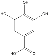
Chemical_Nomenclature : 3,4,5-trihydroxybenzoic acid
Canonical SMILES : C1=C(C=C(C(=C1O)O)O)C(=O)O
InChI : InChI=1S\/C7H6O5\/c8-4-1-3(7(11)12)2-5(9)6(4)10\/h1-2,8-10H,(H,11,12)
InChIKey : LNTHITQWFMADLM-UHFFFAOYSA-N
Other name(s) : Gallic acid,3,4,5-Trihydroxybenzoic acid,Benzoic acid, 3,4,5-trihydroxy-,Gallic acid, tech.
MW : 170.12
Formula : C7H6O5
CAS_number : 149-91-7
PubChem : 370
UniChem : LNTHITQWFMADLM-UHFFFAOYSA-N
IUPHAR :
Wikipedia :
Target
References (4)
| Title : Gallic Acid as a Non-Selective Inhibitor of alpha\/beta-Hydrolase Fold Enzymes Involved in the Inflammatory Process: The Two Sides of the Same Coin - Toyama_2022_Pharmaceutics_14_ |
| Author(s) : Toyama MH , Rogero A , de Moraes LLF , Fernandes GA , da Cruz Costa CR , Belchor MN , De Carli AM , de Oliveira MA |
| Ref : Pharmaceutics , 14 : , 2022 |
| Abstract : Toyama_2022_Pharmaceutics_14_ |
| ESTHER : Toyama_2022_Pharmaceutics_14_ |
| PubMedSearch : Toyama_2022_Pharmaceutics_14_ |
| PubMedID: 35214100 |
| Title : Crystal structure of fungal tannase from Aspergillus niger - Dong_2021_Acta.Crystallogr.D.Struct.Biol_77_267 |
| Author(s) : Dong L , McKinstry WJ , Pan L , Newman J , Ren B |
| Ref : Acta Crystallographica D Struct Biol , 77 :267 , 2021 |
| Abstract : Dong_2021_Acta.Crystallogr.D.Struct.Biol_77_267 |
| ESTHER : Dong_2021_Acta.Crystallogr.D.Struct.Biol_77_267 |
| PubMedSearch : Dong_2021_Acta.Crystallogr.D.Struct.Biol_77_267 |
| PubMedID: 33559614 |
| Gene_locus related to this paper: aspnc-a2qir3 |
| Title : Crystal structure of tannase from lactobacillusplantarum - Ren_2013_J.Mol.Biol_425_2737 |
| Author(s) : Ren B , Wu M , Wang Q , Peng X , Wen H , McKinstry WJ , Chen Q |
| Ref : Journal of Molecular Biology , 425 :2737 , 2013 |
| Abstract : Ren_2013_J.Mol.Biol_425_2737 |
| ESTHER : Ren_2013_J.Mol.Biol_425_2737 |
| PubMedSearch : Ren_2013_J.Mol.Biol_425_2737 |
| PubMedID: 23648840 |
| Gene_locus related to this paper: lacpl-tanL |
| Title : Expression, purification, crystallization and preliminary X-ray analysis of tannase from Lactobacillus plantarum. - Wu_2013_Acta.Crystallogr.Sect.F.Struct.Biol.Cryst.Commun_69_456 |
| Author(s) : Wu M , Peng X , Wen H , Wang Q , Chen Q , McKinstry WJ , Ren B |
| Ref : Acta Crystallographica Sect F Struct Biol Cryst Commun , 69 :456 , 2013 |
| Abstract : Wu_2013_Acta.Crystallogr.Sect.F.Struct.Biol.Cryst.Commun_69_456 |
| ESTHER : Wu_2013_Acta.Crystallogr.Sect.F.Struct.Biol.Cryst.Commun_69_456 |
| PubMedSearch : Wu_2013_Acta.Crystallogr.Sect.F.Struct.Biol.Cryst.Commun_69_456 |
| PubMedID: 23545659 |
| Gene_locus related to this paper: lacpl-tanL |