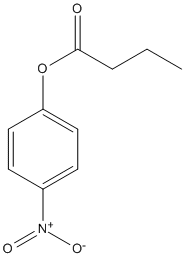
7 structures(e.g. : 7ATF, 5HC2, 5HC0... more)(less)7ATF: Structure of EstD11 in complex with p-Nitrophenol, 5HC2: Structure of esterase Est22 (S188A) with p-nitrophenol, 5HC0: Structure of esterase Est22 with p-nitrophenol, 5AOB: The structure of a novel thermophilic esterase from the Planctomycetes species, Thermogutta terrifontis, Est2-butyrate bound, 3WWC: The complex of pOPH_S172A of pNPB (Pseudomonas sp. Oxidized polyvinyl alcohol hydrolase (OPH)), 3G9U: Crystal structure of EstE5, was soaked by p-nitrophenyl butyrate for 5min, 3G9T: Crystal structure of EstE5, was soaked p-nitrophenyl butyrate for 5sec
The ease with which enzymes can be adapted from their native roles and engineered to function specifically for industrial or commercial applications is crucial to enabling enzyme technology to advance beyond its current state. Directed evolution is a powerful tool for engineering enzymes with improved physical and catalytic properties and can be used to evolve enzymes where lack of structural information may thwart the use of rational design. In this study, we take the versatile and diverse alpha/beta hydrolase fold framework, in the form of dienelactone hydrolase, and evolve it over three unique sequential evolutions with a total of 14 rounds of screening to generate a series of enzyme variants. The native enzyme has a low level of promiscuous activity toward p-nitrophenyl acetate but almost undetectable activity toward larger p-nitrophenyl esters. Using p-nitrophenyl acetate as an evolutionary intermediate, we have generated variants with altered specificity and catalytic activity up to 3 orders of magnitude higher than the native enzyme toward the larger nonphysiological p-nitrophenyl ester substrates. Several variants also possess increased stability resulting from the multidimensional approach to screening. Crystal structure analysis and substrate docking show how the enzyme active site changes over the course of the evolutions as either a direct or an indirect result of mutations.
        
Title: Purification and partial characterization of a novel thermophilic carboxylesterase with high mesophilic specific activity Wood AN, Fernandez-Lafuente R, Cowan DA Ref: Enzyme Microb Technol, 17:816, 1995 : PubMed
An esterase activity obtained from a strain of Bacillus stearothermophilus was purified 5,133-fold to electrophoretic homogeneity with 26% recovery. The purified esterase had a specific activity of 2,032 mumol min-1 mg-1 based on the hydrolysis of p-nitrophenyl caproate at pH 7.0 and 30 degrees C. The apparent molecular mass was 50,000 +/- 2,000 daltons from sodium dodecyl sulfate-polyacrylamide gel electrophoresis and 45,000 +/- 3,000 daltons from gel filtration. Native polyacrylamide gels stained for esterase activity showed three bands. The isoelectric points were estimated to be 5.7, 5.8, and 6.0. Forty amino acid residues were sequenced at the N-terminus. The sequence showed no degeneracy, suggesting that the three esterases are functionally identical carboxylesterases differing by a limited number of amino acids. The enzyme showed maximum activity at pH 7.0 and was very stable at pH 6.0-8.9 with optimum stability at pH 6.0. At this pH and 60 degrees C the half-life was 170 h. Esterase activity was totally inhibited by phenylmethanesulfonyl fluoride, parahydroxymercuribenzoate, eserine, and tosyl-L-phenylalanine, but not by ethylendiaminetetra acetic acid. The esterase obeyed Michaelis-Menten kinetics in the hydrolysis of p-nitrophenyl esters, but both Vmax and KM were protein concentration-dependent. The esterase was able to hydrolyse a number of p-nitrophenyl derivatives (amino acid derivatives and aliphatic acids with different chain lengths).
        
Title: Purification and properties of an Arthrobacter oxydans P52 carbamate hydrolase specific for the herbicide phenmedipham and nucleotide sequence of the corresponding gene Pohlenz HD, Boidol W, Schuttke I, Streber WR Ref: Journal of Bacteriology, 174:6600, 1992 : PubMed
Arthrobacter oxydans P52 isolated from soil samples was found to degrade the phenylcarbamate herbicides phenmedipham and desmedipham cometabolically by hydrolyzing their central carbamate linkages. The phenylcarbamate hydrolase (phenmedipham hydrolase) responsible for the degradative reaction was purified to homogeneity. The enzyme was shown to be a monomer with a molecular weight of 55,000. A 41-kb wild-type plasmid (pHP52) was identified in A. oxydans P52, but not in a derivative of this strain that had spontaneously lost the ability to hydrolyze phenylcarbamates, indicating that the gene for phenylcarbamate degradation (pcd) is plasmid encoded. Determination of two partial amino acid sequences allowed the localization of the coding sequence of the pcd gene on a 3.3-kb PstI restriction fragment within pHP52 DNA by hybridization with synthetic oligonucleotides. The phenylcarbamate hydrolase was functionally expressed in Escherichia coli under control of the lacZ promoter after the 3.3-kb PstI fragment was subcloned into the vector pUC19. A stretch of 1,864 bases within the cloned Pst fragment was sequenced. Sequence analysis revealed an open reading frame of 1,479 bases containing the amino acid partial sequences determined for the purified enzyme. Sequence comparisons revealed significant homology between the pcd gene product and the amino acid sequences of esterases of eukaryotic origin. Subsequently, it was demonstrated that the esterase substrate p-nitrophenylbutyrate is hydrolyzed by phenmedipham hydrolase.
Polyurethanes (PU) are one of the most-used classes of synthetic polymers in Europe, having a considerable impact on the plastic waste management in the European Union. Therefore, they represent a major challenge for the recycling industry, which requires environmentally friendly strategies to be able to re-utilize their monomers without applying hazardous and polluting substances in the process. In this work, enzymatic hydrolysis of a polyurethane-polyester (PU-PE) copolymer using Humicola insolens cutinase (HiC) has been investigated in order to achieve decomposition at milder conditions and avoiding harsh chemicals. PU-PE films have been incubated with the enzyme at 50 C for 168 h, and hydrolysis has been followed throughout the incubation. HiC effectively hydrolysed the polymer, reducing the number average molecular weight (Mn) and the weight average molecular weight (Mw) by 84% and 42%, respectively, as shown by gel permeation chromatography (GPC), while scanning electron microscopy showed cracks at the surface of the PU-PE films as a result of enzymatic surface erosion. Furthermore, Fourier Transform Infrared (FTIR) analysis showed a reduction in the peaks at 1725 cm-1, 1164 cm-1 and 1139 cm-1, indicating that the enzyme preferentially hydrolysed ester bonds, as also supported by the nuclear magnetic resonance spectroscopy (NMR) results. Liquid chromatography time-of-flight/mass spectrometry (LC-MS-Tof) analysis revealed the presence in the incubation supernatant of all of the monomeric constituents of the polymer, thus suggesting that the enzyme was able to hydrolyse both the ester and the urethane bonds of the polymer.
A novel esterase, EstD11, has been discovered in a hot spring metagenomic library. It is a thermophilic and thermostable esterase with an optimum temperature of 60C. A detailed substrate preference analysis of EstD11 was done using a library of chromogenic ester substrate that revealed the broad substrate specificity of EstD11 with significant measurable activity against 16 substrates with varied chain length, steric hindrance, aromaticity and flexibility of the linker between the carboxyl and the alcohol moiety of the ester. The tridimensional structures of EstD11 and the inactive mutant have been determined at atomic resolutions. Structural and bioinformatic analysis, confirm that EstD11 belongs to the family IV, the hormone-sensitive lipase (HSL) family, from the alpha/beta-hydrolase superfamily. The canonical alpha/beta-hydrolase domain is completed by a cap domain, composed by two subdomains that can unmask of the active site to allow the substrate to enter. Eight crystallographic complexes were solved with different substrates and reaction products that allowed identification of the hot-spots in the active site underlying the specificity of the protein. Crystallization and/or incubation of EstD11 at high temperature provided unique information on cap dynamics and a first glimpse of enzymatic activity in vivo. Very interestingly, we have discovered a unique Met zipper lining the active site and the cap domains that could be essential in pivotal aspects as thermo-stability and substrate promiscuity in EstD11
        
Title: Phylogenetic analysis and in-depth characterization of functionally and structurally diverse CE5 cutinases Novy V, Carneiro LV, Shin JH, Larsbrink J, Olsson L Ref: Journal of Biological Chemistry, :101302, 2021 : PubMed
Cutinases are esterases that release fatty acids from the apoplastic layer in plants. As they accept bulky and hydrophobic substrates, cutinases could be used in many applications, ranging from valorization of bark-rich side streams to plastic recycling. Advancement of these applications with cutinases as biocatalysts, however, requires deeper knowledge of the enzymes' biodiversity and structure-function relationships. Here, we mined over 3000 members from Carbohydrate Esterase family 5 (CE5) for putative cutinases and condensed it to 151 genes from known or putative lignocellulose-targeting organisms. The 151 genes were subjected to a phylogenetic analysis. While cutinases with available crystal structures were phylogenetically closely related, we selected nine phylogenic diverse cutinases for characterization. The nine selected cutinases were recombinantly produced and their kinetic activity was characterized against para-nitrophenol substrates esterified with consecutively longer alkyl chains (pNP-C(2) to C(16)). The investigated cutinases each had a unique activity fingerprint against tested pNP-substrates. The five enzymes with the highest activity on pNP-C(12) and C(16), indicative of activity on bulky hydrophobic compounds, were selected for in-depth kinetic and structure-function analysis. All five enzymes showed a decrease in k(cat) values with increasing substrate chain length, while K(M) values and binding energies (calculated from in silico docking analysis) improved. Two cutinases from Fusarium solani and Cryptococcus sp. exhibited outstandingly low K(M) values, resulting in high catalytic efficiencies towards pNP-C(16). Docking analysis suggested that different clades of the phylogenetic tree may harbor enzymes with different modes of substrate interaction, involving a solvent-exposed catalytic triad, a lipase-like lid, or a clamshell-like active site possibly formed by flexible loops.
        
Title: Structure-guided engineering of a Thermobifida fusca cutinase for enhanced hydrolysis on natural polyester substrate Dong Q, Yuan S, Wu L, Su L, Zhao Q, Wu J, Huang W, Zhou J Ref: Bioresour. Bioprocess, 7:37, 2020 : PubMed
Cutinases could degrade insoluble polyester, including natural cutin and synthetic plastic. However, their turnover efficiency for polyester remains too low for industrial application. Herein, we report the 1.54-A resolution X-ray crystal structure of a cutinase from Thermobifida fusca and modeling structure in complex with a cutin mimic oligo-polyester C24H42O8. These efforts subsequently guided our design of cutinase variants with less bulky residues in the vicinity of the substrate binding site. The L90A and I213A variants exhibit increased hydrolysis activity (5- and 2.4-fold, respectively) toward cutin and also showed enhanced cotton scouring efficiency compared with the wild-type enzyme.
Hormone sensitive lipase (HSL) catalyzes the hydrolysis of triacylglycerols into fatty acids and glycerol, thus playing key roles in energy homeostasis. However, the application of HSL serving as a pharmaceutical target and an industrial biocatalyst is largely hampered due to the lack of high-resolution structural information. Here we report biochemical properties and crystal structures of a novel HSL homologue esterase Est22 from a deep-sea metagenomic library. Est22 prefers short acyl chain esters and has a very high activity with substrate p-nitrophenyl butyrate. The crystal structures of wild type and mutated Est22 with its product p-nitrophenol are solved with resolutions ranging from 1.4 A to 2.43 A. The Est22 exhibits a alpha/beta-hydrolase fold consisting with a catalytic domain and a substrate-recognizing cap domain. Residues Ser188, Asp287, and His317 comprise the catalytic triad in the catalytic domain. The p-nitrophenol molecule occupies the substrate binding pocket and forms hydrogen bonds with adjacent residues Gly108, Gly109, and Gly189. Est22 exhibits a dimeric form in solution, whereas mutants D287A and H317A change to polymeric form, which totally abolished its enzymatic activities. Our study provides insights into the catalytic mechanism of HSL family esterase and facilitates the understanding for further industrial and biotechnological applications of esterases.
        
Title: Characterization of LipN (Rv2970c) of Mycobacterium Tuberculosis H37Rv and its Probable Role in Xenobiotic Degradation Jadeja D, Dogra N, Arya S, Singh G, Kaur J Ref: Journal of Cellular Biochemistry, 117:390, 2016 : PubMed
LipN (Rv2970c) belongs to the Lip family of M. tuberculosis H37Rv and is homologous to the human Hormone Sensitive Lipase. The enzyme demonstrated preference for short carbon chain substrates with optimal activity at 45 degrees C/pH 8.0 and stability between pH 6.0-9.0. The specific activity of the enzyme was 217 U/mg protein with pNP-butyrate as substrate. It hydrolyzed tributyrin to di- and monobutyrin. The active-site residues of the enzyme were confirmed to be Ser216, Asp316, and His346. Tetrahydrolipstatin, RHC-80267 and N-bromosuccinimide inhibited LipN enzyme activity completely. Interestingly, Trp145, a non active-site residue, demonstrated functional role to retain enzyme activity. The enzyme was localized in cytosolic fraction of M. tuberculosis H37Rv. The enzyme was able to synthesize ester of butyric acid, methyl butyrate, in presence of methanol. LipN was able to hydrolyze 4-hydroxyphenylacetate to hydroquinone. The gene was not expressed in in-vitro growth conditions while the expression of rv2970c gene was observed post 6h of macrophage infection by M. tuberculosis H37Ra. Under individual in-vitro stress conditions, the gene was expressed during acidic stress condition only. These findings suggested that LipN is a cytosolic, acid inducible carboxylesterase with no positional specificity in demonstrating activity with short carbon chain substrates. It requires Trp145, a non active site residue, for it's enzyme activity. J. Cell. Biochem. 117: 390-401, 2016. (c) 2015 Wiley Periodicals, Inc.
Diacylglycerol lipase alpha (DAGLalpha) hydrolyses DAG to generate the principal endocannabinoid (eCB) 2-arachidonoylglycerol (2-AG) in the central nervous system. DAGLalpha dependent cannabinoid (CB) signalling has been implicated in numerous processes including axonal growth and guidance, adult neurogenesis and retrograde signalling at the synapse. Recent studies have implicated DAGLalpha as an emerging drug target for several conditions including pain and obesity. Activity assays are critical to the drug discovery process; however, measurement of diacylglycerol lipase (DAGL) activity using its native substrate generally involves low-throughput MS techniques. Some relatively high-throughput membrane based assays utilizing surrogate substrates have been reported, but these do not take into account the rate-limiting effects often associated with the ability of a drug to cross the cell membrane. In the present study, we report the development of a live cell assay to measure DAGLalpha activity. Two previously reported DAGLalpha surrogate substrates, p-nitrophenyl butyrate (PNPB) and 6,8-difluoro-4-methylumbelliferyl octanoate (DiFMUO), were evaluated for their ability to detect DAGLalpha activity in live cell assays using a human cell line stably expressing the human DAGLalpha transgene. Following optimization, the small molecule chromogenic substrate PNPB proved to be superior by providing lower background activity along with a larger signal window between transfected and parental cells when compared with the fluorogenic substrate DiFMUO. The assay was further validated using established DAGL inhibitors. In summary, the live cell DAGLalpha assay reported here offers an economical and convenient format to screen for novel inhibitors as part of drug discovery programmes and compliments previously reported high-throughput membrane based DAGL assays.
        
Title: Directed evolution of new and improved enzyme functions using an evolutionary intermediate and multidirectional search Porter JL, Boon PL, Murray TP, Huber T, Collyer CA, Ollis DL Ref: ACS Chemical Biology, 10:611, 2015 : PubMed
The ease with which enzymes can be adapted from their native roles and engineered to function specifically for industrial or commercial applications is crucial to enabling enzyme technology to advance beyond its current state. Directed evolution is a powerful tool for engineering enzymes with improved physical and catalytic properties and can be used to evolve enzymes where lack of structural information may thwart the use of rational design. In this study, we take the versatile and diverse alpha/beta hydrolase fold framework, in the form of dienelactone hydrolase, and evolve it over three unique sequential evolutions with a total of 14 rounds of screening to generate a series of enzyme variants. The native enzyme has a low level of promiscuous activity toward p-nitrophenyl acetate but almost undetectable activity toward larger p-nitrophenyl esters. Using p-nitrophenyl acetate as an evolutionary intermediate, we have generated variants with altered specificity and catalytic activity up to 3 orders of magnitude higher than the native enzyme toward the larger nonphysiological p-nitrophenyl ester substrates. Several variants also possess increased stability resulting from the multidimensional approach to screening. Crystal structure analysis and substrate docking show how the enzyme active site changes over the course of the evolutions as either a direct or an indirect result of mutations.
        
Title: Roles of tryptophan residue and disulfide bond in the variable lid region of oxidized polyvinyl alcohol hydrolase Yang Y, Ko TP, Liu L, Li J, Huang CH, Chen J, Guo RT, Du G Ref: Biochemical & Biophysical Research Communications, 452:509, 2014 : PubMed
Oxidized polyvinyl alcohol hydrolase (OPH) catalyzes the cleavage of C-C bond in beta-diketone. It belongs to the alpha/beta-hydrolase family and contains a unique lid region that covers the active site. The lid is the most variable region when pOPH from Pseudomonas sp. VM15C and sOPH from Sphingopyxis sp. 113P3 are compared. The wild-type enzymes and the pOPH mutants W255A, W255Y and W255F were analyzed for lipase activity by using p-nitrophenyl (pNP) esters as the substrates. The wild-type enzymes showed increased Km and decreased kcat/Km with the acyl chain length, and the mutants showed reduced kcat/Km for pNP acetate, indicating the importance of Trp255 in sequestering the active site from solvent. The significantly lower activity for pNP butyrate can be a result of product inhibition, as suggested by the complex crystal structures, in which butyric acid, DMSO or PEG occupied the same substrate-binding cleft. The mutant activity was retained with pNP caprylate and pNP laurate as the substrates, reflecting the amphipathic nature of the cleft. Moreover, the disulfide bond formation of Cys257/267 is important for the activity of pOPH, but it is not essential for sOPH, which has a shorter lid structure.
        
Title: SulE, a sulfonylurea herbicide de-esterification esterase from Hansschlegelia zhihuaiae S113 Hang BJ, Hong Q, Xie XT, Huang X, Wang CH, He J, Li SP Ref: Applied Environmental Microbiology, 78:1962, 2012 : PubMed
De-esterification is an important degradation or detoxification mechanism of sulfonylurea herbicide in microbes and plants. However, the biochemical and molecular mechanisms of sulfonylurea herbicide de-esterification are still unknown. In this study, a novel esterase gene, sulE, responsible for sulfonylurea herbicide de-esterification, was cloned from Hansschlegelia zhihuaiae S113. The gene contained an open reading frame of 1,194 bp, and a putative signal peptide at the N terminal was identified with a predicted cleavage site between Ala37 and Glu38, resulting in a 361-residue mature protein. SulE minus the signal peptide was synthesized in Escherichia coli BL21 and purified to homogeneity. SulE catalyzed the de-esterification of a variety of sulfonylurea herbicides that gave rise to the corresponding herbicidally inactive parent acid and exhibited the highest catalytic efficiency toward thifensulfuron-methyl. SulE was a dimer without the requirement of a cofactor. The activity of the enzyme was completely inhibited by Ag(+), Cd(2+), Zn(2+), methamidophos, and sodium dodecyl sulfate. A sulE-disrupted mutant strain, DeltasulE, was constructed by insertion mutation. DeltasulE lost the de-esterification ability and was more sensitive to the herbicides than the wild type of strain S113, suggesting that sulE played a vital role in the sulfonylurea herbicide resistance of the strain. The transfer of sulE into Saccharomyces cerevisiae BY4741 conferred on it the ability to de-esterify sulfonylurea herbicides and increased its resistance to the herbicides. This study has provided an excellent candidate for the mechanistic study of sulfonylurea herbicide metabolism and detoxification through de-esterification, construction of sulfonylurea herbicide-resistant transgenic crops, and bioremediation of sulfonylurea herbicide-contaminated environments.
        
Title: Non-lipolytic and lipolytic sequence-related carboxylesterases: a comparative study of the structure-function relationships of rabbit liver esterase 1 and bovine pancreatic bile-salt-activated lipase Chahinian H, Fantini J, Garmy N, Manco G, Sarda L Ref: Biochimica & Biophysica Acta, 1801:1195, 2010 : PubMed
To differentiate esterases from lipases at the structure-function level, we have compared the kinetic properties and structural features of sequence-related esterase 1 from rabbit liver (rLE) and bile-salt-activated lipase from bovine pancreas (bBAL). In contrast to rLE, bBAL hydrolyses water-insoluble medium and long chain esters as vinyl laurate, trioctanoin and olive oil. Conversely, rLE and bBAL are both active on water-soluble short chain esters as vinyl acetate, vinyl propionate, vinyl butyrate, tripropionin, tributyrin and p-nitrophenyl butyrate. However, the enzymes show distinctive kinetic behaviours. rLE displays maximal activity at low substrate concentration, below the critical micelle concentration, whereas bBAL acts preferencially on emulsified esters, at concentration exceeding the solubility limit. Comparison of the 3D structures of rLE and bBAL shows, in particular, that the peptide loop at positions 116-123 in bBAL is deleted in rLE. This peptide segment interacts with a bile salt molecule thus inducing a conformational transition which gives access to the active site. Inhibition studies and manual docking of a bulky ester molecule as vinyl laurate in the catalytic pocket of rLE and bBAL show that the inability of the esterase to hydrolyse large water-insoluble esters is not due to steric hindrance. It is hypothesized that esterases lack specific hydrophobic structures involved both in the stabilization of the lipase-lipid adsorption complex at interfaces and in the spontaneous transfer of a single substrate molecule from interface to the catalytic site.
The bacterial thermoalkalophilic lipases that hydrolyze saturated fatty acids at 60-75 degrees C and pH 8-10 are grouped as the lipase family I.5. We report here the crystal structure of the lipase from Geobacillus thermocatenulatus, the first structure of a member of the lipase family I.5 showing an open configuration. Unexpectedly, enzyme activation involves large structural rearrangements of around 70 amino acids and the concerted movement of two lids, the alpha6- and alpha7-helices, unmasking the active site. Central in the restructuring process of the lids are both the transfer of bulky hydrophobic residues out of the N-terminal end of the alpha6-helix and the incorporation of short side chain residues to the alpha6 C-terminal end. All these structural changes are stabilized by the Zn(2+)-binding domain, which is characteristic of this family of lipases. Two detergent molecules are placed in the active site, mimicking chains of the triglyceride substrate, demonstrating the position of the oxyanion hole and the three pockets that accommodate the sn-1, sn-2, and sn-3 fatty acids chains. The combination of structural and biochemical studies indicate that the lid opening is not mediated by temperature but triggered by interaction with lipid substrate.
In all sequenced genomes, a large fraction of predicted genes encodes proteins of unknown biochemical function and up to 15% of the genes with "known" function are mis-annotated. Several global approaches are routinely employed to predict function, including sophisticated sequence analysis, gene expression, protein interaction, and protein structure. In the first coupling of genomics and enzymology, Phizicky and colleagues undertook a screen for specific enzymes using large pools of partially purified proteins and specific enzymatic assays. Here we present an overview of the further developments of this approach, which involve the use of general enzymatic assays to screen individually purified proteins for enzymatic activity. The assays have relaxed substrate specificity and are designed to identify the subclass or sub-subclasses of enzymes (phosphatase, phosphodiesterase/nuclease, protease, esterase, dehydrogenase, and oxidase) to which the unknown protein belongs. Further biochemical characterization of proteins can be facilitated by the application of secondary screens with natural substrates (substrate profiling). We demonstrate here the feasibility and merits of this approach for hydrolases and oxidoreductases, two very broad and important classes of enzymes. Application of general enzymatic screens and substrate profiling can greatly speed up the identification of biochemical function of unknown proteins and the experimental verification of functional predictions produced by other functional genomics approaches.
        
Title: Cloning and characterization of EstC from Burkholderia gladioli, a novel-type esterase related to plant enzymes Reiter B, Glieder A, Talker D, Schwab H Ref: Applied Microbiology & Biotechnology, 54:778, 2000 : PubMed
By screening a genomic library of Burkholderia gladioli (formerly Pseudomonas marginata) for clones exhibiting esterolytic activity, the gene for a novel-type esterase (EstC) showing significant homology to plant enzymes could be isolated. High homology was found to two hydroxynitrile lyases originating from Hevea brasiliensis (tropical rubber tree) and Manihot esculenta (cassava), and to two proteins from Oryza sativa (rice) that are specifically induced upon infection by Pseudomonas syringae pv. syringae. The sequenced ORF encodes for a protein of 298 amino acids. The enzyme was efficiently overexpressed in Escherichia coli, purified and characterized with respect to enzymatic capabilities. The enzyme was able to hydrolyze a variety of esterase substrates of low to medium carbonic acid chain length, but no triglycerides were hydrolyzed. Despite the high sequence homology, no hydroxynitrile lyase activity could be recognized.
        
Title: Purification and partial characterization of a novel thermophilic carboxylesterase with high mesophilic specific activity Wood AN, Fernandez-Lafuente R, Cowan DA Ref: Enzyme Microb Technol, 17:816, 1995 : PubMed
An esterase activity obtained from a strain of Bacillus stearothermophilus was purified 5,133-fold to electrophoretic homogeneity with 26% recovery. The purified esterase had a specific activity of 2,032 mumol min-1 mg-1 based on the hydrolysis of p-nitrophenyl caproate at pH 7.0 and 30 degrees C. The apparent molecular mass was 50,000 +/- 2,000 daltons from sodium dodecyl sulfate-polyacrylamide gel electrophoresis and 45,000 +/- 3,000 daltons from gel filtration. Native polyacrylamide gels stained for esterase activity showed three bands. The isoelectric points were estimated to be 5.7, 5.8, and 6.0. Forty amino acid residues were sequenced at the N-terminus. The sequence showed no degeneracy, suggesting that the three esterases are functionally identical carboxylesterases differing by a limited number of amino acids. The enzyme showed maximum activity at pH 7.0 and was very stable at pH 6.0-8.9 with optimum stability at pH 6.0. At this pH and 60 degrees C the half-life was 170 h. Esterase activity was totally inhibited by phenylmethanesulfonyl fluoride, parahydroxymercuribenzoate, eserine, and tosyl-L-phenylalanine, but not by ethylendiaminetetra acetic acid. The esterase obeyed Michaelis-Menten kinetics in the hydrolysis of p-nitrophenyl esters, but both Vmax and KM were protein concentration-dependent. The esterase was able to hydrolyse a number of p-nitrophenyl derivatives (amino acid derivatives and aliphatic acids with different chain lengths).
        
Title: Purification and properties of an Arthrobacter oxydans P52 carbamate hydrolase specific for the herbicide phenmedipham and nucleotide sequence of the corresponding gene Pohlenz HD, Boidol W, Schuttke I, Streber WR Ref: Journal of Bacteriology, 174:6600, 1992 : PubMed
Arthrobacter oxydans P52 isolated from soil samples was found to degrade the phenylcarbamate herbicides phenmedipham and desmedipham cometabolically by hydrolyzing their central carbamate linkages. The phenylcarbamate hydrolase (phenmedipham hydrolase) responsible for the degradative reaction was purified to homogeneity. The enzyme was shown to be a monomer with a molecular weight of 55,000. A 41-kb wild-type plasmid (pHP52) was identified in A. oxydans P52, but not in a derivative of this strain that had spontaneously lost the ability to hydrolyze phenylcarbamates, indicating that the gene for phenylcarbamate degradation (pcd) is plasmid encoded. Determination of two partial amino acid sequences allowed the localization of the coding sequence of the pcd gene on a 3.3-kb PstI restriction fragment within pHP52 DNA by hybridization with synthetic oligonucleotides. The phenylcarbamate hydrolase was functionally expressed in Escherichia coli under control of the lacZ promoter after the 3.3-kb PstI fragment was subcloned into the vector pUC19. A stretch of 1,864 bases within the cloned Pst fragment was sequenced. Sequence analysis revealed an open reading frame of 1,479 bases containing the amino acid partial sequences determined for the purified enzyme. Sequence comparisons revealed significant homology between the pcd gene product and the amino acid sequences of esterases of eukaryotic origin. Subsequently, it was demonstrated that the esterase substrate p-nitrophenylbutyrate is hydrolyzed by phenmedipham hydrolase.