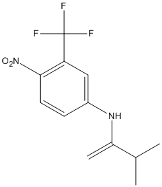
Flutamide and its more potent active metabolite 2-hydroxyflutamide competitively block dihydrotestosterone binding at androgen receptors, forming inactive complexes which cannot translocate into the cell nucleus. Formation of inactive receptors inhibits androgen-dependent DNA and protein synthesis, resulting in tumor cell growth arrest or transient tumor regression. (NCI04). Flutamide has been linked to numerous cases of acute liver injury, which are frequently severe and can be fatal.
Flutamide, an antiandrogen drug, is widely used for the treatment of prostate cancer. The initial metabolic pathways of flutamide are hydroxylation and hydrolysis. It was recently reported that the hydrolyzed product, 4-nitro-3-(trifluoromethyl)phenylamine (FLU-1), is further metabolized to N-hydroxy FLU-1, an assumed hepatotoxicant. However, the esterase responsible for the flutamide hydrolysis has not been characterized. In the present study, we found that human arylacetamide deacetylase (AADAC) efficiently hydrolyzed flutamide using recombinant AADAC expressed in COS7 cells. In contrast, carboxylesterase1 (CES1) and CES2, which are responsible for the hydrolysis of many drugs, could not hydrolyze flutamide. AADAC is specifically expressed in the endoplasmic reticulum. Flutamide hydrolase activity was highly detected in human liver microsomes (K(m), 794 +/- 83 microM; V(max), 1.1 +/- 0.0 nmol/min/mg protein), whereas the activity was extremely low in human liver cytosol. The flutamide hydrolase activity in human liver microsomes was strongly inhibited by bis-(p-nitrophenyl)phosphate [corrected], diisopropylphosphorofluoride, and physostigmine sulfate (eserine) but moderately inhibited by sodium fluoride, phenylmethylsulfonyl fluoride, and disulfiram. The same inhibition pattern was obtained with the recombinant AADAC. Moreover, human liver and jejunum microsomes showing AADAC expression could hydrolyze flutamide, but human pulmonary and renal microsomes, which do not express AADAC, showed slight activity. In human liver microsomal samples (n = 50), the flutamide hydrolase activities were significantly correlated with the expression levels of AADAC protein (r = 0.66, p < 0.001). In conclusion, these results clearly showed that flutamide is exclusively hydrolyzed by AADAC. AADAC would be an important enzyme responsible for flutamide-induced hepatotoxicity.
        
Title: Detection of a new N-oxidized metabolite of flutamide, N-[4-nitro-3-(trifluoromethyl)phenyl]hydroxylamine, in human liver microsomes and urine of prostate cancer patients Goda R, Nagai D, Akiyama Y, Nishikawa K, Ikemoto I, Aizawa Y, Nagata K, Yamazoe Y Ref: Drug Metabolism & Disposition: The Biological Fate of Chemicals, 34:828, 2006 : PubMed
Flutamide (2-methyl-N-[4-nitro-3-(trifluoromethyl)phenyl]-propanamide), a nonsteroidal antiandrogen, is used in the treatment of prostate cancer but is occasionally associated with hepatic dysfunction. In the present study, the metabolism of flutamide including the formation of the possible reactive toxic metabolites was investigated using human liver microsomes and 10 isoforms of recombinant human cytochrome P450 (P450). 2-Hydroxyflutamide (OH-flutamide) and 4-nitro-3-(trifluoromethyl)phenylamine (FLU-1) were the main products of flutamide metabolism in human liver microsomes. The formation of OH-flutamide was markedly inhibited by ellipticine, an inhibitor of CYP1A1/1A2, and was mainly catalyzed by the recombinant CYP1A2. FLU-1 was also produced from OH-flutamide, but its metabolic rate was much less than that from flutamide. An inhibitor of carboxylesterase, bis-(p-nitrophenyl)phosphoric acid, completely inhibited the formation of FLU-1 from flutamide in human liver microsomes. A new metabolite, N-[4-nitro-3-(trifluoromethyl)phenyl]hydroxylamine (FLU-1-N-OH), was detected as a product of the reaction of FLU-1 with human liver microsomes and identified by comparison with the synthetic standard. The formation of FLU-1-N-OH was markedly inhibited by the addition of miconazole, an inhibitor of CYP3A4, and was mediated by recombinant CYP3A4. Furthermore, FLU-1-N-OH was detected mostly as the conjugates (glucuronide/sulfate) in the urine of prostate cancer patients collected for 3 h after treatment with flutamide. The formation of FLU-1-N-OH, however, did not differ between patients with and without abnormalities of hepatic functions among a total of 29 patients. The lack of an apparent association of the urinary excretion of FLU-1-N-OH and hepatic disorder may suggest the involvement of an additional unknown factor in the mechanisms of flutamide hepatotoxicity.
        
Title: [Changes in liver function induced by flutamide in patients with prostate cancer (studies in patients treated with total androgen blockage)] Ikemoto I, Ohishi Y, Yamazaki H, Wada T, Aizawa Y Ref: Nihon Hinyokika Gakkai Zasshi, 91:556, 2000 : PubMed
OBJECTIVES: The incidence of flutamide-induced liver toxicity was studied in 30 consecutive patients with prostate cancer who were treated with total androgen blockage (TAB) therapy (luteinizing hormone releasing hormone [LHRH] analogue and flutamide) in our hospital during the last 3 years and in 20 consecutive patients with prostate cancer who were treated by partial androgen blockage (PAB) therapy (LHRH analogue alone). METHODS: Liver function test, including measurement of serum levels of aspartate aminotransferase (AST) alanine aminotransferase (ALT), total cholesterol, total bilirubin, gamma-glutamyl transpeptidase (gamma-GTP), and cholinesterase were performed at regular interval. RESULTS: The incidence of liver toxicity in patients receiving TAB (10 cases of 25 patients) was significantly higher than in patients receiving PAB (2 of 18 patients). Two patients in whom severe liver toxicity developed after receiving TAB were hospitalized. However, after flutamide was discontinued all patients with liver damage recovered with normalization of AST and ALT levels. Levels of total cholesterol and gamma-GTP did not differ significantly in either patient group. In two patients receiving TAB total bilirubin levels showed slight, transient elevations after maximum elevations of AST and ALT. In 80% of patients receiving TAB serum levels of cholinesterase were significantly higher than those in patients receiving PAB. CONCLUSION: These data suggest that the risk of flutamide-induced liver toxicity is significant in patients receiving TAB. However, this damage can be normalized after flutamide has been discontinued. Serum levels of cholinesterase also increase significantly in patients receiving TAB. This previously unreported phenomenon suggests an unknown effect of flutamide on liver function in patients with prostate cancer.
        
1 lessTitle: Contributions of arylacetamide deacetylase and carboxylesterase 2 to flutamide hydrolysis in human liver Kobayashi Y, Fukami T, Shimizu M, Nakajima M, Yokoi T Ref: Drug Metabolism & Disposition: The Biological Fate of Chemicals, 40:1080, 2012 : PubMed
Flutamide, an antiandrogen drug, is widely used for the treatment of prostate cancer. The major metabolic pathways of flutamide are hydroxylation and hydrolysis. The hydrolyzed metabolite, 5-amino-2-nitrobenzotrifluoride (FLU-1), is further metabolized to N-hydroxy FLU-1, an assumed hepatotoxicant. Our previous study demonstrated that arylacetamide deacetylase (AADAC), one of the major serine esterases expressed in the human liver and gastrointestinal tract, catalyzes the flutamide hydrolysis. However, the enzyme kinetics in human tissue microsomes were not consistent with the kinetics by recombinant human AADAC. Thus, it seemed that AADAC is not the sole enzyme responsible for flutamide hydrolysis in human. In the present study, we found that recombinant carboxylesterase (CES) 2 could hydrolyze flutamide at low concentrations of flutamide. In the inhibition assay, the flutamide hydrolase activities at a flutamide concentration of 5 muM in human liver and jejunum microsomes were strongly inhibited by a selective CES2 inhibitor, 10 muM loperamide, with the residual activities of 22.9 +/- 3.5 and 18.6 +/- 0.7%, respectively. These results suggest that CES2 is also involved in the flutamide hydrolysis in human tissues. Using six individual human livers, the contributions of AADAC and CES2 to flutamide hydrolysis were estimated by using the relative activity factor. The relative contribution of CES2 was approximately 75 to 99% at the concentration of 5 muM flutamide. In contrast, the relative contribution of AADAC increased in parallel with the concentration of flutamide. Thus, CES2, rather than AADAC, largely contributed to the flutamide hydrolysis in clinical therapeutics.
        
Title: Human arylacetamide deacetylase is a principal enzyme in flutamide hydrolysis Watanabe A, Fukami T, Nakajima M, Takamiya M, Aoki Y, Yokoi T Ref: Drug Metabolism & Disposition: The Biological Fate of Chemicals, 37:1513, 2009 : PubMed
Flutamide, an antiandrogen drug, is widely used for the treatment of prostate cancer. The initial metabolic pathways of flutamide are hydroxylation and hydrolysis. It was recently reported that the hydrolyzed product, 4-nitro-3-(trifluoromethyl)phenylamine (FLU-1), is further metabolized to N-hydroxy FLU-1, an assumed hepatotoxicant. However, the esterase responsible for the flutamide hydrolysis has not been characterized. In the present study, we found that human arylacetamide deacetylase (AADAC) efficiently hydrolyzed flutamide using recombinant AADAC expressed in COS7 cells. In contrast, carboxylesterase1 (CES1) and CES2, which are responsible for the hydrolysis of many drugs, could not hydrolyze flutamide. AADAC is specifically expressed in the endoplasmic reticulum. Flutamide hydrolase activity was highly detected in human liver microsomes (K(m), 794 +/- 83 microM; V(max), 1.1 +/- 0.0 nmol/min/mg protein), whereas the activity was extremely low in human liver cytosol. The flutamide hydrolase activity in human liver microsomes was strongly inhibited by bis-(p-nitrophenyl)phosphate [corrected], diisopropylphosphorofluoride, and physostigmine sulfate (eserine) but moderately inhibited by sodium fluoride, phenylmethylsulfonyl fluoride, and disulfiram. The same inhibition pattern was obtained with the recombinant AADAC. Moreover, human liver and jejunum microsomes showing AADAC expression could hydrolyze flutamide, but human pulmonary and renal microsomes, which do not express AADAC, showed slight activity. In human liver microsomal samples (n = 50), the flutamide hydrolase activities were significantly correlated with the expression levels of AADAC protein (r = 0.66, p < 0.001). In conclusion, these results clearly showed that flutamide is exclusively hydrolyzed by AADAC. AADAC would be an important enzyme responsible for flutamide-induced hepatotoxicity.
        
Title: Detection of a new N-oxidized metabolite of flutamide, N-[4-nitro-3-(trifluoromethyl)phenyl]hydroxylamine, in human liver microsomes and urine of prostate cancer patients Goda R, Nagai D, Akiyama Y, Nishikawa K, Ikemoto I, Aizawa Y, Nagata K, Yamazoe Y Ref: Drug Metabolism & Disposition: The Biological Fate of Chemicals, 34:828, 2006 : PubMed
Flutamide (2-methyl-N-[4-nitro-3-(trifluoromethyl)phenyl]-propanamide), a nonsteroidal antiandrogen, is used in the treatment of prostate cancer but is occasionally associated with hepatic dysfunction. In the present study, the metabolism of flutamide including the formation of the possible reactive toxic metabolites was investigated using human liver microsomes and 10 isoforms of recombinant human cytochrome P450 (P450). 2-Hydroxyflutamide (OH-flutamide) and 4-nitro-3-(trifluoromethyl)phenylamine (FLU-1) were the main products of flutamide metabolism in human liver microsomes. The formation of OH-flutamide was markedly inhibited by ellipticine, an inhibitor of CYP1A1/1A2, and was mainly catalyzed by the recombinant CYP1A2. FLU-1 was also produced from OH-flutamide, but its metabolic rate was much less than that from flutamide. An inhibitor of carboxylesterase, bis-(p-nitrophenyl)phosphoric acid, completely inhibited the formation of FLU-1 from flutamide in human liver microsomes. A new metabolite, N-[4-nitro-3-(trifluoromethyl)phenyl]hydroxylamine (FLU-1-N-OH), was detected as a product of the reaction of FLU-1 with human liver microsomes and identified by comparison with the synthetic standard. The formation of FLU-1-N-OH was markedly inhibited by the addition of miconazole, an inhibitor of CYP3A4, and was mediated by recombinant CYP3A4. Furthermore, FLU-1-N-OH was detected mostly as the conjugates (glucuronide/sulfate) in the urine of prostate cancer patients collected for 3 h after treatment with flutamide. The formation of FLU-1-N-OH, however, did not differ between patients with and without abnormalities of hepatic functions among a total of 29 patients. The lack of an apparent association of the urinary excretion of FLU-1-N-OH and hepatic disorder may suggest the involvement of an additional unknown factor in the mechanisms of flutamide hepatotoxicity.
        
Title: [Changes in liver function induced by flutamide in patients with prostate cancer (studies in patients treated with total androgen blockage)] Ikemoto I, Ohishi Y, Yamazaki H, Wada T, Aizawa Y Ref: Nihon Hinyokika Gakkai Zasshi, 91:556, 2000 : PubMed
OBJECTIVES: The incidence of flutamide-induced liver toxicity was studied in 30 consecutive patients with prostate cancer who were treated with total androgen blockage (TAB) therapy (luteinizing hormone releasing hormone [LHRH] analogue and flutamide) in our hospital during the last 3 years and in 20 consecutive patients with prostate cancer who were treated by partial androgen blockage (PAB) therapy (LHRH analogue alone). METHODS: Liver function test, including measurement of serum levels of aspartate aminotransferase (AST) alanine aminotransferase (ALT), total cholesterol, total bilirubin, gamma-glutamyl transpeptidase (gamma-GTP), and cholinesterase were performed at regular interval. RESULTS: The incidence of liver toxicity in patients receiving TAB (10 cases of 25 patients) was significantly higher than in patients receiving PAB (2 of 18 patients). Two patients in whom severe liver toxicity developed after receiving TAB were hospitalized. However, after flutamide was discontinued all patients with liver damage recovered with normalization of AST and ALT levels. Levels of total cholesterol and gamma-GTP did not differ significantly in either patient group. In two patients receiving TAB total bilirubin levels showed slight, transient elevations after maximum elevations of AST and ALT. In 80% of patients receiving TAB serum levels of cholinesterase were significantly higher than those in patients receiving PAB. CONCLUSION: These data suggest that the risk of flutamide-induced liver toxicity is significant in patients receiving TAB. However, this damage can be normalized after flutamide has been discontinued. Serum levels of cholinesterase also increase significantly in patients receiving TAB. This previously unreported phenomenon suggests an unknown effect of flutamide on liver function in patients with prostate cancer.