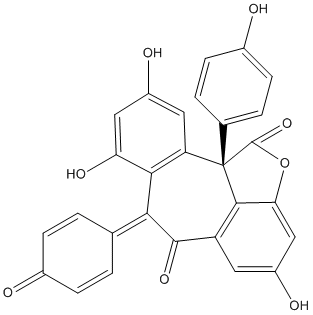
Phytochemical investigation of the stem wood of Dipterocarpus alatus led to the isolation and characterization of four new oligostilbenoids, dipterocarpols A-D (1-4), together with two known resveratrol oligomers, hopeahainol (5) and hopeafuran (6). The structures of the new compounds were determined by comprehensive spectral analysis including 1D and 2D NMR, and high-resolution MS. The absolute configurations were determined by NOESY and CD spectra. Dipterocarpol A (1) and hopeahainol A (5) showed moderate acetylcholinesterase inhibitory activity with IC50 values of 8.28 microM and 11.28 microM, respectively. Furthermore, the discovery of compound 3 gave the first evidence that the biosynthetic origin of resveratrol aneuploids is related to the loss of a half resveratrol unit by oxidative cleavage.
Bioassay-guided investigation of the stem bark of Hopea chinensis led to the isolation of two new polyphenols, hopeachinols C(1) and D(2), together with ten known compounds (3-12). Compounds 1 and 2 were determined by extensive analysis of spectroscopic data and computational methods. All of these phytochemicals were tested for acetylcholinesterase inhibitory activity, and five resveratrol-derived compounds (1 and 7-10) exhibited significant activity with their IC(5)(0) values ranging from 4.81 to 11.71 microM.
A phytochemical study of Hopea hainanensis has led to the isolation of three new polyphenols and one known compound. The most important of these compounds are hopeahainols A (2) and B (3), which contain an unprecedented carbon skeleton. The structures were elucidated by analysis of the spectroscopic data including single-crystal X-ray spectroscopy and computational methods. Hopeahainol A was an acetylcholinesterase inhibitor with an IC50 value of 4.33 microM, which is comparable to that of huperzine A, a presently prescribed drug for the treatment of Alzheimer, while other similar structures were inactive. This observation was complemented by a 3D interaction model of the inhibitor with active sites.
        
6 lessTitle: Hopeahainol A binds reversibly at the acetylcholinesterase (AChE) peripheral site and inhibits enzyme activity with a novel higher order concentration dependence Rosenberry TL, Martin PK, Nix AJ, Wildman SA, Cheung J, Snyder SA, Tan RX Ref: Chemico-Biological Interactions, 259:78, 2016 : PubMed
Natural product inhibitors of AChE are of interest both because they offer promise as inexpensive drugs for symptomatic relief in Alzheimer's disease and because they may provide insights into the structural features of the AChE catalytic site. Hopeahainol A is an uncharged polyphenol AChE inhibitor from the stem bark of H. hainanensis with a constrained, partially dearomatized bicyclic core. Molecular modeling indicates that hopeahainol A binds at the entrance of the long but narrow AChE active site gorge because it is too bulky to be accommodated within the gorge without severe distortion of the gorge as depicted in AChE crystal structures. We conducted inhibitor competition experiments in which AChE inhibition was measured with hopeahainol A together with either edrophonium (which binds at the base of the gorge) or thioflavin T (which binds to the peripheral or P-site near the gorge mouth). The results agreed with the molecular modeling and indicated that hopeahainol A at lower concentrations (<200 muM) bound only to the P-site, as hopeahainol A and thioflavin T were unable to form a ternary complex with AChE while hopeahainol A and edrophonium did form a ternary complex with essentially no competition between them. Inhibition increased to a striking extent at higher concentrations of hopeahainol A, with plots analogous to classic Dixon plots showing a dependence on hopeahainol A concentrations to the third- or fourth order. The inhibition at higher hopeahainol A concentrations was completely reversed on dilution and blocked by bound edrophonium. We hypothesize that bound hopeahainol A induces conformational changes in the AChE active site that allow binding of additional hopeahainol A molecules, a phenomenon that would be unprecedented for a reversible inhibitor that apparently forms no covalent bonds with AChE.
Phytochemical investigation of the stem wood of Dipterocarpus alatus led to the isolation and characterization of four new oligostilbenoids, dipterocarpols A-D (1-4), together with two known resveratrol oligomers, hopeahainol (5) and hopeafuran (6). The structures of the new compounds were determined by comprehensive spectral analysis including 1D and 2D NMR, and high-resolution MS. The absolute configurations were determined by NOESY and CD spectra. Dipterocarpol A (1) and hopeahainol A (5) showed moderate acetylcholinesterase inhibitory activity with IC50 values of 8.28 microM and 11.28 microM, respectively. Furthermore, the discovery of compound 3 gave the first evidence that the biosynthetic origin of resveratrol aneuploids is related to the loss of a half resveratrol unit by oxidative cleavage.
Increasing evidence demonstrates that amyloid beta (Abeta) elicits mitochondrial dysfunction and oxidative stress, which contributes to the pathogenesis of Alzheimer's disease (AD). Identification of the molecules targeting Abeta is thus of particular significance in the treatment of AD. Hopeahainol A (HopA), a polyphenol with a novel skeleton obtained from Hopea hainanensis, is potentially acetylcholinesterase-inhibitory and anti-oxidative in H(2) O(2) -treated PC12 cells. In this study, we reported that HopA might bind to Abeta(1-42) directly and inhibit the Abeta(1-42) aggregation using a combination of molecular dynamics simulation, binding assay, transmission electron microscopic analysis and staining technique. We also demonstrated that HopA decreased the interaction between Abeta(1-42) and Abeta-binding alcohol dehydrogenase, which in turn reduced mitochondrial dysfunction and oxidative stress in vivo and in vitro. In addition, HopA was able to rescue the long-term potentiation induction by protecting synaptic function and attenuate memory deficits in APP/PS1 mice. Our data suggest that HopA might be a promising drug for therapeutic intervention in AD.
        
Title: Total syntheses of hopeanol and hopeahainol A empowered by a chiral Bronsted acid induced pinacol rearrangement Snyder SA, Thomas SB, Mayer AC, Breazzano SP Ref: Angew Chem Int Ed Engl, 51:4080, 2012 : PubMed
Bioassay-guided investigation of the stem bark of Hopea chinensis led to the isolation of two new polyphenols, hopeachinols C(1) and D(2), together with ten known compounds (3-12). Compounds 1 and 2 were determined by extensive analysis of spectroscopic data and computational methods. All of these phytochemicals were tested for acetylcholinesterase inhibitory activity, and five resveratrol-derived compounds (1 and 7-10) exhibited significant activity with their IC(5)(0) values ranging from 4.81 to 11.71 microM.
Title: Total synthesis and biological evaluation of the resveratrol-derived polyphenol natural products hopeanol and hopeahainol A Nicolaou KC, Kang Q, Wu TR, Lim CS, Chen DY Ref: Journal of the American Chemical Society, 132:7540, 2010 : PubMed
The total synthesis and biological evaluation of the resveratrol-derived natural products hopeanol (2) and hopeahainol A (3) in their racemic and antipodal forms are described. The Friedel-Crafts-based synthetic strategy employed was developed from model studies that established the feasibility of constructing the C(7b) quaternary center through an intramolecular Friedel-Crafts reaction and a Grob-type fragmentation to introduce an obligatory olefinic bond in the growing molecule. The final stages of the synthesis involved an epoxide substrate and an intramolecular Friedel-Crafts reaction, followed by oxidation to afford, upon global deprotection, hopeahainol A (3). The latter was converted under basic conditions to hopeanol (2), whereas the reverse transformation, previously suggested as a step in the biosynthesis of hopeahainol A (3), was not observed under a variety of conditions. Biological evaluation of the synthesized compounds confirmed the reported acetylcholinesterase inhibitory activity of hopeahainol A (3) but not the reported cytotoxic potencies of hopeanol (2).
        
Title: Protective effect of hopeahainol A, a novel acetylcholinesterase inhibitor, on hydrogen peroxide-induced injury in PC12 cells Shi da H, Wu JH, Ge HM, Tan RX Ref: Environ Toxicol Pharmacol, 28:30, 2009 : PubMed
In this study, we evaluated the effects of hopeahainol A, a novel acetylcholinesterase inhibitor (AChEI) from Hopea hainanensis, on H(2)O(2)-induced cytotoxicity in PC12 cells and the possible mechanism. Exposure of PC12 cells to 200muM H(2)O(2) caused cell apoptosis, reduction in cell viability and antioxidant enzyme activities, increment in malondialdehyde (MDA) level, and leakage of lactate dehydrogenase (LDH). Pretreatment of the cells with hopeahainol A at 0.1-10muM before H(2)O(2) exposure significantly attenuated those changes in a dose-dependent manner. Moreover, hopeahainol A could mitigate intracellular accumulation of reactive oxygen species (ROS) and Ca(2+), the loss of mitochondrial membrane potential (MMP), and the increase of caspase-3, -8 and -9 activities induced by H(2)O(2). These results show that hopeahainol A protects PC12 cells from H(2)O(2) injury by modulating endogenous antioxidant enzymes, scavenging ROS and prevention of apoptosis. There was potential for hopeahainol A to be used in treating Alzheimer's disease (AD) that involved acetylcholinesterase, free radical, oxidative damage and cell apoptosis.
A phytochemical study of Hopea hainanensis has led to the isolation of three new polyphenols and one known compound. The most important of these compounds are hopeahainols A (2) and B (3), which contain an unprecedented carbon skeleton. The structures were elucidated by analysis of the spectroscopic data including single-crystal X-ray spectroscopy and computational methods. Hopeahainol A was an acetylcholinesterase inhibitor with an IC50 value of 4.33 microM, which is comparable to that of huperzine A, a presently prescribed drug for the treatment of Alzheimer, while other similar structures were inactive. This observation was complemented by a 3D interaction model of the inhibitor with active sites.