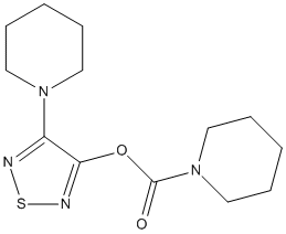
Apart from Lysosomal acid lipase human-LIPA, Lalistat-1 and -2 also inhibit major cytosolic lipid hydrolases responsible for lipid degradation in primary cells at neutral pH through off-target effects (ATGL not alpha/beta hydrolas; HSL hormone sensitive lipase); MGL Monoglyceride lipase). Lalistat also impairs Mycobacterium tuberculosis growth by targeting hydolases LipR Rv3084 Rv1984c Rv0183
OBJECTIVES: Lysosomal acid lipase (LAL) is the key enzyme, which degrades neutral lipids at an acidic pH in lysosomes. The role of LAL in various cellular processes has mostly been studied in LAL-knockout mice, which share phenotypical characteristics with humans suffering from LAL deficiency. In vitro, the cell-specific functions of LAL have been commonly investigated by using the LAL inhibitors Lalistat-1 and Lalistat-2. METHODS: We performed lipid hydrolase activity assays and serine hydrolase-specific activity-based labeling combined with quantitative proteomics to investigate potential off-target effects of Lalistat-1 and -2. RESULTS: Pharmacological LAL inhibition but not genetic loss of LAL impairs isoproterenol-stimulated lipolysis as well as neutral triglyceride and cholesteryl ester hydrolase activities. Apart from LAL, Lalistat-1 and -2 also inhibit major cytosolic lipid hydrolases responsible for lipid degradation in primary cells at neutral pH through off-target effects. Their binding to the active center of the enzymes leads to a decrease in neutral lipid hydrolase activities in cells overexpressing the respective enzymes. CONCLUSIONS: Our findings are critically important since they demonstrate that commonly used concentrations of these inhibitors are not suitable to investigate the role of LAL-specific lipolysis in lysosomal function, signaling pathways, and autophagy. The interpretation of their effects on lipid metabolism should be taken with caution and the applied inhibitor concentrations in cell culture studies should not exceed 1 microM.
Lysosomal acid lipase (LAL) is essential for cholesteryl ester (CE) and triacylglycerol (TAG) hydrolysis in lysosomes. Clinically, an autosomal recessive LIPA mutation causes LAL deficiency (LAL-D), either Wolman Disease or Cholesterol Ester Storage Disease (CESD). LAL-D is associated with ectopic neutral lipid accumulation in the liver, small intestine, spleen, adrenal glands, and blood. Considering the importance of unesterified cholesterol and fatty acids in bone metabolism, we hypothesized that LAL is essential to bone formation, and ultimately, skeletal health. To investigate the role of LAL in skeletal homeostasis, we used LAL-deficient ((-/-)) mice and osteoblast cell cultures. Male LAL(-/-) mice had lower trabecular BV/TV (12%) compared to WT mice (21%), due to decreased trabecular number and increased trabecular separation; this change was not apparent in the females. While both sexes of LAL(-/-) mice displayed decreased cortical bone thickness and polar moment of inertia, only the female LAL(-/-) mice showed increased cortical porosity. Histological analyses revealed that LAL(-/-) mice tended to have less osteoblasts but no change in osteoclast numbers. In studying the cell-autonomous role of LAL, we observed impaired osteoblastogenesis of LAL(-/-) calvarial osteoblasts and in bone marrow stromal cells treated with the LAL inhibitor lalistat. Consistent with LAL's role in other tissues, lalistat resulted in profound lipid puncta accumulation and an altered intracellular lipid profile. Finally, we analyzed a large de-identified national insurance database (i.e. 2016/2017 Optum Clinformatics(a)) which revealed that adults (<=18 years) with CESD (n=3,076) had a higher odds ratio (OR=1.21; 95% CI=1.03-1.41) of all-cause fracture at any location compared to adults without CESD (n=13.7 M) after adjusting for demographic variables and osteoporosis. These data demonstrate that alterations in LAL have significant clinical implications related to fracture risk and that LAL's modulation of lipid metabolism is a critical for osteoblast function.
        
Title: Low levels of Lysosomal Acid Lipase (LAL) activity increases necroinflammation in adult patients with biopsy-proven metabolic associated fatty liver disease Thoen RU, Longo L, Neto SC, Alvares-da-Silva MR Ref: Clin Res Hepatol Gastroenterol, 45:101638, 2021 : PubMed
INTRODUCTION AND OBJECTIVE: Metabolic associated fatty liver disease (MAFLD), characterized by intra-hepatic fat accumulation, will soon be the leading cause of end-stage liver disease. Lysosomal Acid Lipase (LAL) is a key enzyme in lipid metabolism. We investigated its activity in patients with biopsy-proven MAFLD. METHODS: Prospective cross-sectional study in patients with biopsy-proven MAFLD. Blood LAL-activity (pmol/punch/h) was measured with dried blood spot extracts using Lalistat 2. Demographic, clinical, and laboratory data were collected. RESULTS: 101 adult patients were recruited. Among them, 11.9% had a diagnosis of MAFLD without steatohepatitis and 88.1% had MAFLD with steatohepatitis. The median of LAL-activity in patients with MAFLD was 76.8 pmol/punch/h. MAFLD patients with steatohepatitis showed an increase in gamma-glutamyl transferase (p = 0.042), insulin (p = 0.001), homeostatic model assessment for insulin resistance (HOMA-IR, p = 0.001) and advanced liver fibrosis (p < 0.001), compared to cases of MAFLD without steatohepatitis. There was no statistical difference in LAL-activity between the cases (p = 0.296). When considering LAL-activity above and below 77 pmol/punch/h as a cut-off value, patients with reduced LAL-activity had a significant increase in necroinflammatory activity according to the METAVIR score (p = 0.040), and NAFLD activity score (NAS, p = 0.031) compared to cases with higher LAL-activity. CONCLUSION: Our findings suggest that reduced LAL-activity is associated with increased necroinflammatory activity and severity of the NAS. A better knowledge of the role of LAL may provide new insights into the pathogenesis and progression of MAFLD.
Therapy-induced senescence in cancer cells is an irreversible antiproliferative state, which inhibits tumor growth and is therefore a potent anti-neoplastic mechanism. In this study, low doses of Abrus agglutinin (AGG)-induced senescence through autophagy in prostate carcinoma cells (PC3) and inhibited proliferation. The inhibition of autophagy with 3-methyl adenine reversed AGG-induced senescence, thus confirming that AGG-triggered senescence required autophagy. AGG treatment also led to lipophagy-mediated accumulation of free fatty acids (FFAs), with a concomitant decrease in the number of lipid droplets. Lalistat, a lysosomal acid lipase inhibitor, abrogated AGG-induced lipophagy and senescence in PC3 cells, indicating that lipophagy is essential for AGG-induced senescence. The accumulation of FFAs increased reactive oxygen species generation, a known facilitator of senescence, which was also reduced in the presence of lalistat. Furthermore, AGG upregulated silent mating type information regulator 2 homolog 1 (SIRT1), while the presence of sirtinol reduced autophagy flux and the senescent phenotype in the AGG-treated cells. Mechanistically, AGG-induced cytoplasmic SIRT1 deacetylated a Lys residue on the cytoplasmic domain of lysosome-associated membrane protein 1 (LAMP1), an autolysosomal protein, resulting in lipophagy and senescence. Taken together, our findings demonstrate a novel SIRT1/LAMP1/lipophagy axis mediating AGG-induced senescence in prostate cancer cells.
        
Title: Lysosomal acid lipase is the major acid retinyl ester hydrolase in cultured human hepatic stellate cells but not essential for retinyl ester degradation Wagner C, Hois V, Pajed L, Pusch LM, Wolinski H, Trauner M, Zimmermann R, Taschler U, Lass A Ref: Biochimica & Biophysica Acta Molecular & Cellular Biology Lipids, 1865:158730, 2020 : PubMed
Vitamin A is stored as retinyl esters (REs) in lipid droplets of hepatic stellate cells (HSCs). To date, two different pathways are known to facilitate the breakdown of REs: (i) Hydrolysis of REs by neutral lipases, and (ii) whole lipid droplet degradation in autolysosomes by acid hydrolysis. In this study, we evaluated the contribution of neutral and acid RE hydrolases to the breakdown of REs in human HSCs. (R)-Bromoenol lactone (R-BEL), inhibitor of adipose triglyceride lipase (ATGL) and patatin-like phospholipase domain-containing 3 (PNPLA3), the hormone-sensitive lipase (HSL) inhibitor 76-0079, as well as the serine-hydrolase inhibitor Orlistat reduced neutral RE hydrolase activity of LX-2 cell-lysates between 20 and 50%. Interestingly, in pulse-chase experiments, R-BEL, 76-0079, as well as Orlistat exerted little to no effect on cellular RE breakdown of LX-2 cells as well as primary human HSCs. In contrast, Lalistat2, a specific lysosomal acid lipase (LAL) inhibitor, virtually blunted acid in vitro RE hydrolase activity of LX-2 cells. Accordingly, HSCs isolated from LAL-deficient mice showed RE accumulation and were virtually devoid of acidic RE hydrolase activity. In pulse-chase experiments however, LAL-deficient HSCs, similar to LX-2 cells and primary human HSCs, were not defective in degrading REs. In summary, results demonstrate that ATGL, PNPLA3, and HSL contribute to neutral RE hydrolysis of human HSCs. LAL is the major acid RE hydrolase in HSCs. Yet, LAL is not limiting for RE degradation under serum-starvation. Together, results suggest that RE breakdown of HSCs is facilitated by (a) so far unknown, non-Orlistat inhibitable RE-hydrolase(s).
Laboratory diagnostics of lysosomal acid lipase deficiency (LAL-D), a rare disorder associated with LIPA alterations, are based on the evaluation of LAL activity. In dry blood spots (DBS) submitted for LAL-D diagnostics (the screening cohort) over a two-year period or obtained from a cohort of retrospective LAL-D patients, we measured: (1) LAL activity using a two-reaction assay with 4-methylumbelliferone palmitate (4-MU-Palm) and Lalistat-2, a specific LAL inactivator; (2) total lipase (TL) activity by a 1-hour kinetic 4-MU-Palm cleavage reaction (no Lalistat-2). The TL activity was expressed as the area under the kinetic curve after 1 hour (TL-AUC1h) of the reaction and presented as the median (min-max). LAL activity was reduced in 30/537 individuals from the screening cohort, among which LIPA sequencing revealed six patients and one carrier. Overall, 16 (89%) individuals among six novel and 12 retrospective LAL-D patients carried at least one c.894G>A mutation (six were homozygous). The TL-AUC1h in nonLAL-D specimens with normal LAL activity (n = 90) was unambiguously higher (9471 [4015-23 585] RFU*h/punch) compared to LAL-D patients, including six new and nine retrospective patients (1810 [357-2608] RFU*h/punch). Importantly, in 13/15 examined nonLAL-D specimens with reduced LAL activity the TL-AUC1h was above a threshold of 2652 RFU*h/punch. Applying this threshold, the TL-AUC1h index discriminated all LAL-D patients (100% sensitivity) and 103/105 nonLAL-D specimens (98% specificity). Given that there is no need for Lalistat-2 and two parallel enzymatic reactions in conjunction with high sensitivity and specificity, the kinetic assay seems to be practical for LAL-D screening. Synopsis: Lysosomal acid lipase deficiency responsible for Wolman disease and cholesterol ester storage disease could be reliably detected using a kinetic assay of total lipase activity with a fluorogenic substrate.
BACKGROUND: Deficiency of lysosomal acid lipase (LAL) causes Wolman disease and cholesterol ester storage disease. With the recent introduction of enzyme replacement therapy to manage LAL deficiency comes the need for a reliable assay of LAL enzymatic activity that can be applied to dried blood spots (DBS). METHODS: We prepared and tested a library of analogs of palmitoyl 4-methylumbelifferyl esters to find a highly active and specific substrate for LAL in DBS. The LAL assay was optimized leading to both LC-MS/MS and fluorometric assay of LAL. We tested the new assay on DBS from healthy and LAL-deficient patients. RESULTS: The ester formed between palmitic acid and 4-propyl-8-methyl-7-hydroxycoumarin (P-PMHC) was found to be >98% selective for LAL in DBS based on the sensitivity of its activity to the LAL-specific inactivator Lalistat-2 and the fact that the activity was close to zero using DBS from patients previously shown to be LAL-deficient. Use of P-PMHC and heavy isotope-labeled internal standard with optimized assay conditions led to an approximately 2-fold increase in the specific activity of LAL compared with the previously reported LAL assay. Patients deficient in LAL were readily distinguished from normal persons with the new LAL assay using UPLC-MS/MS or fluorometric assay platforms. CONCLUSIONS: The new assay can measure LAL in DBS with a single measurement compared with the previous method involving 2 assays done in parallel.
        
Title: Best practice in the measurement and interpretation of lysosomal acid lipase in dried blood spots using the inhibitor Lalistat 2 Lukacs Z, Barr M, Hamilton J Ref: Clinica Chimica Acta, 471:201, 2017 : PubMed
Lysosomal acid lipase deficiency (LAL-D) is an inherited, autosomal recessive lysosomal storage disorder characterized by progressive damage in multiple organ systems. Diagnosis is especially important in infants, in whom the course of disease is rapidly lethal without treatment. The recent regulatory approval of recombinant human lysosomal acid lipase (LAL), sebelipase alfa, merits rapid diagnosis in clinical routine, particularly in infants. A method for measuring LAL activity in dried blood spot (DBS) samples using the highly specific LAL inhibitor Lalistat 2 is available. This method is shown to effectively discriminate between individuals with LAL-D and unaffected controls. With the increase in DBS LAL testing since the original publication of this method, a need to optimise assay performance has been identified. Here, we describe refinements to the DBS assay, including technical modifications, quality control measures and best-practice guidance for interpreting and reporting results. Particular attention is paid to alternatives to the use of mercuric chloride as the stop reagent and the choice of excitation wavelength for 4-methylumbelliferone palmitate under assay conditions at pH4.0. In addition, a simpler method of reporting results is proposed using cutoffs based on percentage mean normal enzyme activity.
        
Title: Human lysosomal acid lipase inhibitor lalistat impairs Mycobacterium tuberculosis growth by targeting bacterial hydrolases Davda D, Martin BR Ref: Medchemcomm, 7:1797, 2016 : PubMed
Lalistat inhibits growth of Mycobacterium tuberculosis in bacterial culture as well as in infected macrophages. Target identification by quantitative proteomics revealed a cluster of 20 hydrolytic proteins including members of the lipase family. Lipases are essential for M. tuberculosis fatty acid production and energy
storage thus representing promising antibiotic targets.
        
Title: Extended use of a selective inhibitor of acid lipase for the diagnosis of Wolman disease and cholesteryl ester storage disease Civallero G, De Mari J, Bittar C, Burin M, Giugliani R Ref: Gene, 539:154, 2014 : PubMed
Lysosomal acid lipase (LAL) deficiency produces two well defined inborn disorders, Wolman disease (WD) and cholesteryl ester storage disease (CESD). WD is a severe, early-onset condition involving massive storage of triglycerides and cholesteryl esters in the liver, with death usually occurring before one year of life. CESD is a more attenuated, later-onset disease that leads to a progressive and variable liver dysfunction. Diagnosis of LAL deficiency is mainly based on the enzyme assay of LAL activity in fibroblasts. Recently, a selective acid lipase inhibitor was used for the determination of enzyme activity in dried-blood filter paper (DBFP) samples. To extend and to validate these studies, we tested LAL activity with selective inhibition on DBFP samples, leukocytes and fibroblasts. Our results showed a clear discrimination between patients with LAL deficiency and healthy controls when using DBFP, leukocytes or fibroblasts (p<0.001). Deficiency of LAL was also demonstrated in individuals referred to our laboratory with suspected clinical diagnosis of WD, CESD, and Niemann-Pick type B. We conclude that the assay of LAL using selective inhibitor is a reliable and useful method for the identification of LAL deficiency, not only in DBFP samples but also in leukocytes and fibroblasts. This is important as enzyme replacement therapy for LAL deficiency is currently being developed, making the correct diagnosis a critical issue.
        
Title: A practical fluorometric assay method to measure lysosomal acid lipase activity in dried blood spots for the screening of cholesteryl ester storage disease and Wolman disease Dairaku T, Iwamoto T, Nishimura M, Endo M, Ohashi T, Eto Y Ref: Mol Genet Metab, 111:193, 2014 : PubMed
Fluorometric measurements of 4-methylumbelliferone (4-MU) are generally used to screen lysosomal storage diseases (LSDs) using dried blood spots (DBSs). However, in DBS, it is difficult to measure lysosomal acid lipase (LAL) activity due to the influence of other lipases in whole blood. Recently, Hamilton used a fluorometric enzyme assay with 4-MU derivatives to measure the LAL activity in DBS. This method requires mercury chloride as stopping reagent, and the fluorescence intensity of 4-MU was measured at an acidic pH. We report a revised method to measure the LAL activity without using toxic mercury chloride and to measure the fluorescence intensity of 4-MU at a basic pH. For this measurement, we established a more practical method that does not require mercury chloride. The LAL activity in DBS was measured in 51 normal controls, seven obligate carriers and seven patients with CESD. The average LAL activities +/- SD in the DBS from the normal, obligate carriers and CESD patients were 0.68 +/- 0.2 (range: 0.3-1.08), 0.21 +/- 0.1 (range: 0.11-0.41) and 0.02 +/- 0.02 (range: 0-0.06) nmol/punch/h, respectively. There was a significant difference between the normal and the CESD. Our method does not require toxic mercury chloride and is an appropriate revised enzyme assay using DBS for screening patients with CESD.
        
Title: A new method for the measurement of lysosomal acid lipase in dried blood spots using the inhibitor Lalistat 2 Hamilton J, Jones I, Srivastava R, Galloway P Ref: Clinica Chimica Acta, 413:1207, 2012 : PubMed
BACKGROUND: Cholesterol ester storage disease (CESD) and Wolman Disease (WD) are due to deficiency of lysosomal acid lipase (LAL). A new method is described for the measurement of LAL in dried blood spots (DBS) using Lalistat 2 an inhibitor of LAL. METHODS: LAL activity in DBS extracts was measured using the substrate 4-methylumbelliferyl palmitate. LAL activity was determined by measuring total lipase activity and lipase activity in the presence of Lalistat 2. The specificity of Lalistat 2 was investigated using human recombinant LAL (hrLAL) and human pancreatic lipase (hPL). RESULTS: Lalistat 2 inhibited hrLAL with 1% residual activity at 1 muM inhibitor but had no effect on hPL up to 10 muM. LAL activity in DBS samples obtained from normal controls (n=140) was 0.50-2.30 nmol/punch/h and in patients with CESD was <0.03 nmol/punch/h (n=11). Activity in carriers showed intermediate activity: 0.15-0.40 nmol/punch/h (n=15). CONCLUSIONS: Measurement of LAL using DBS is made difficult by the presence of other lipases in whole blood. Lalistat 2 is a specific inhibitor of LAL which allows the determination of LAL in DBS. Results show the method differentiates clearly between normal controls, carriers and affected cases.