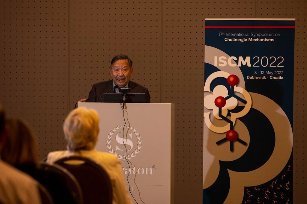
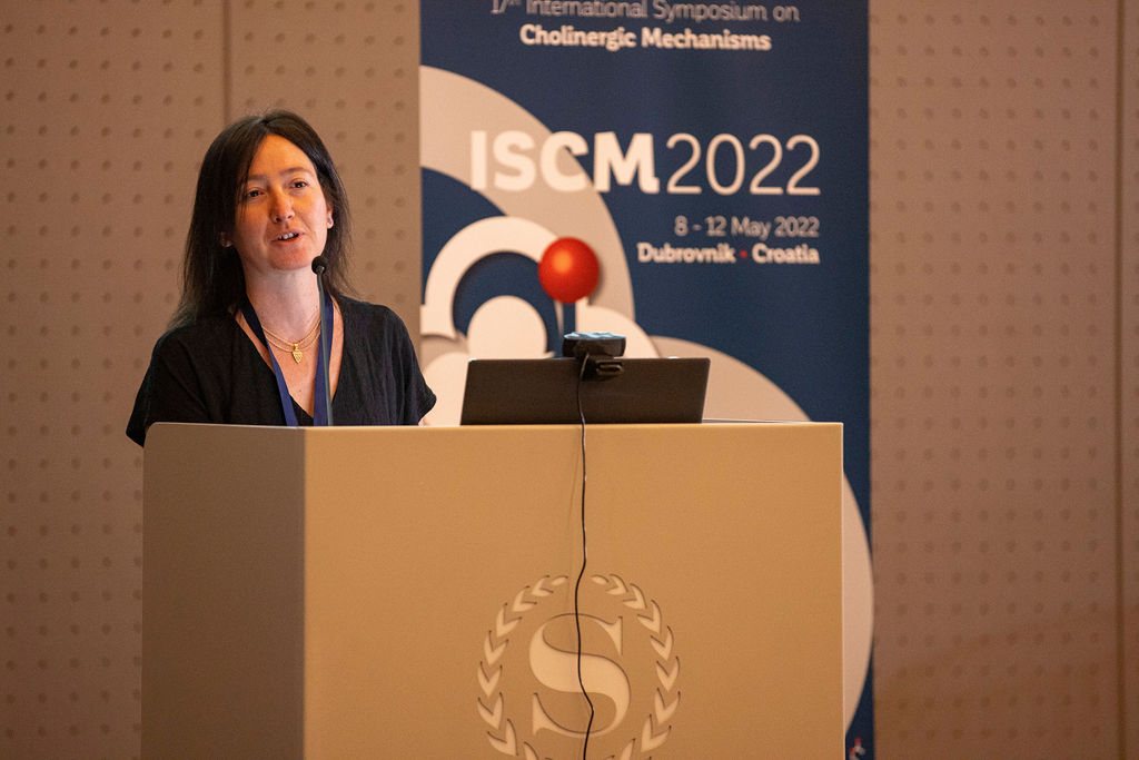
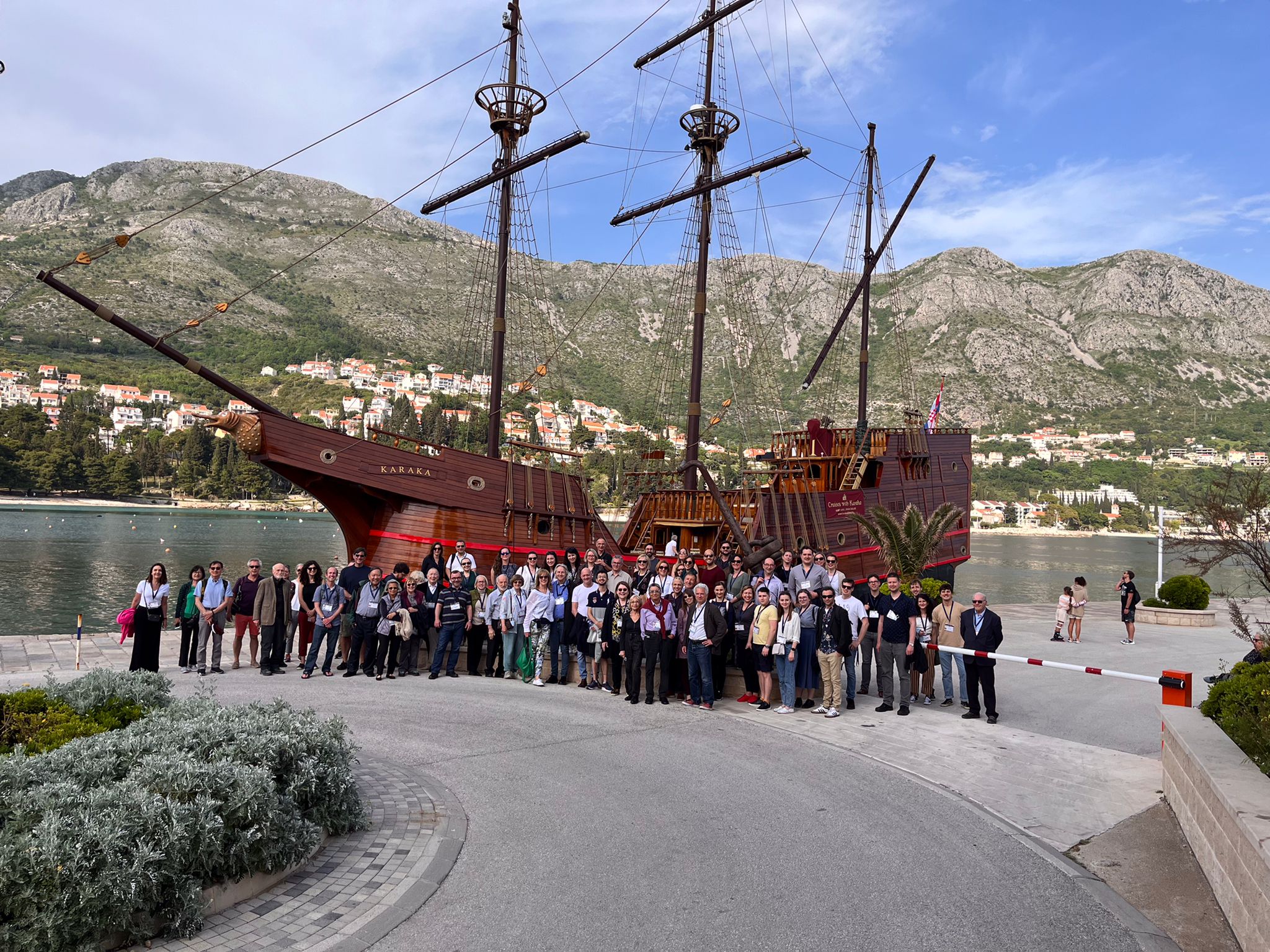
The 17th International Symposium on Cholinergic Mechanisms
2022 Dubrovnik , Croatia
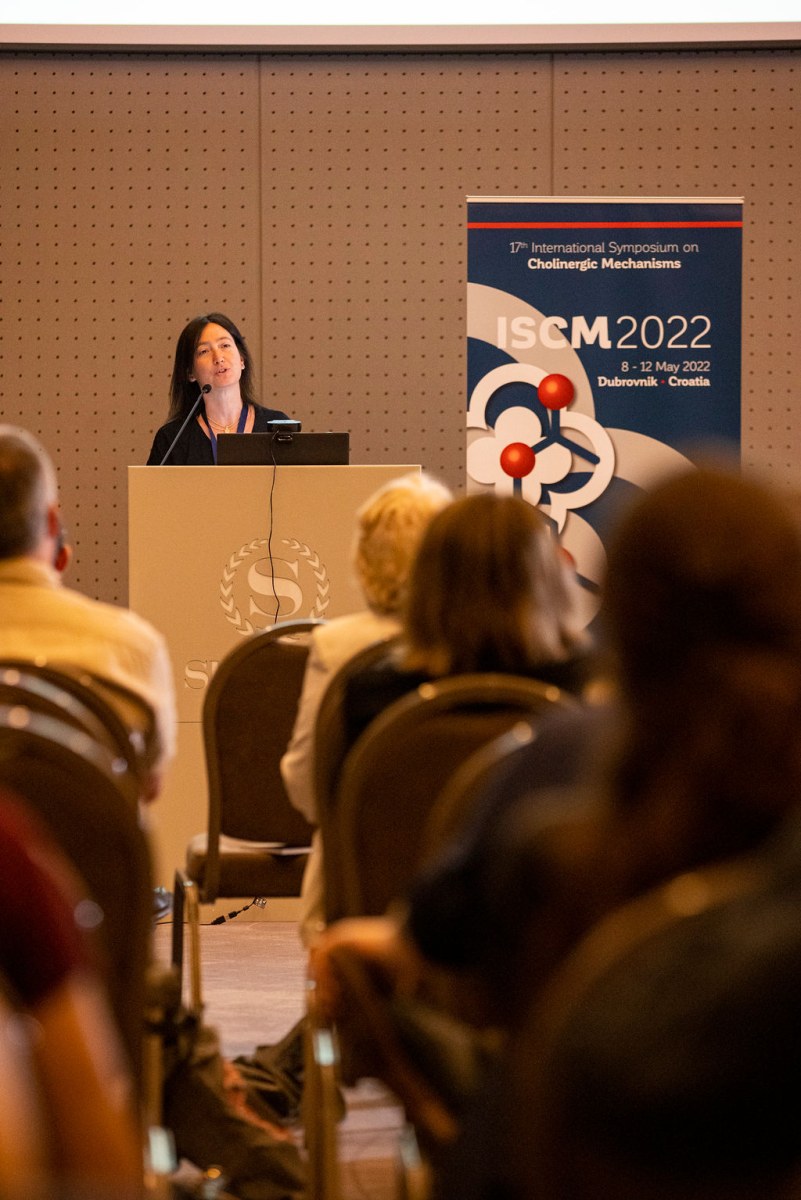
The 17th International Symposium on Cholinergic Mechanisms
Proceedings:
The 17th International Symposium on Cholinergic Mechanisms
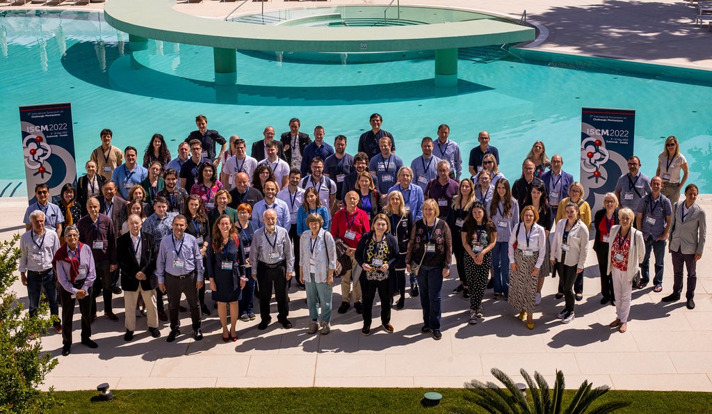
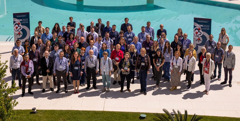
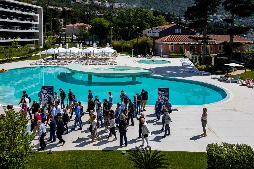
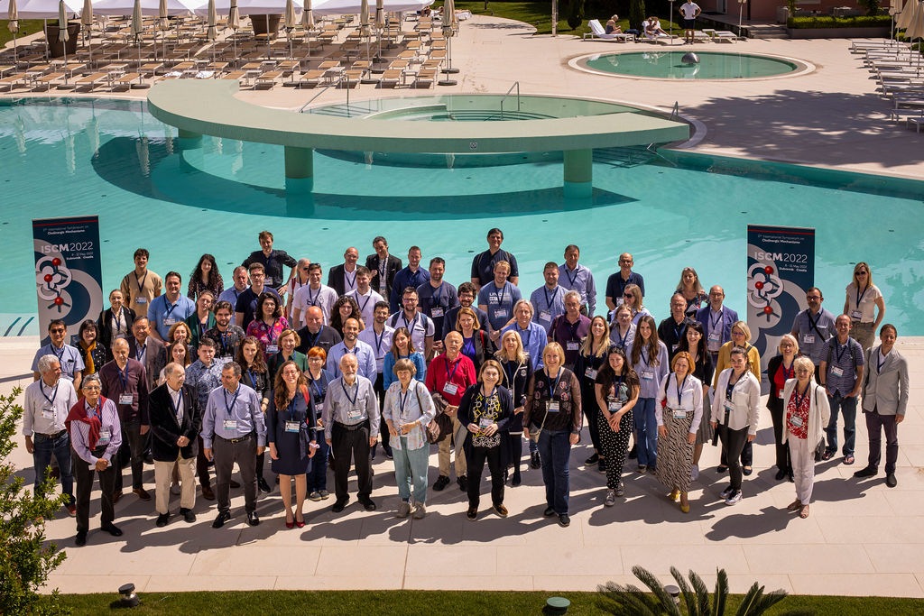
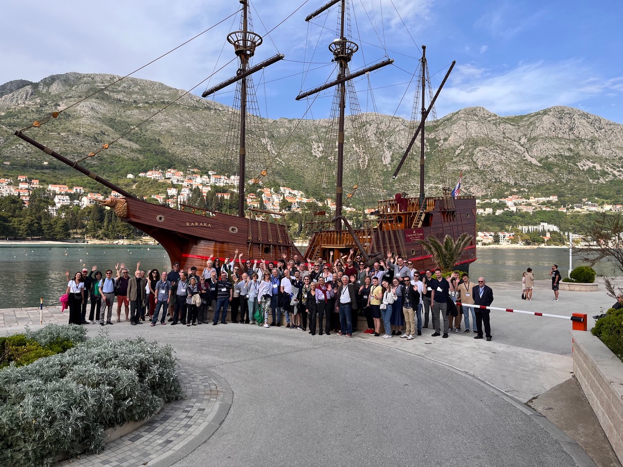
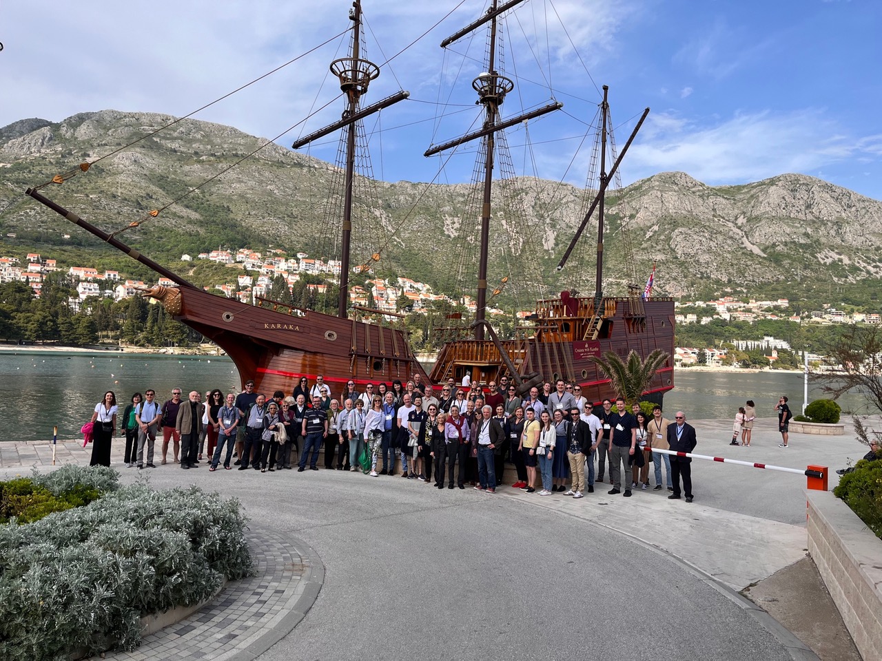
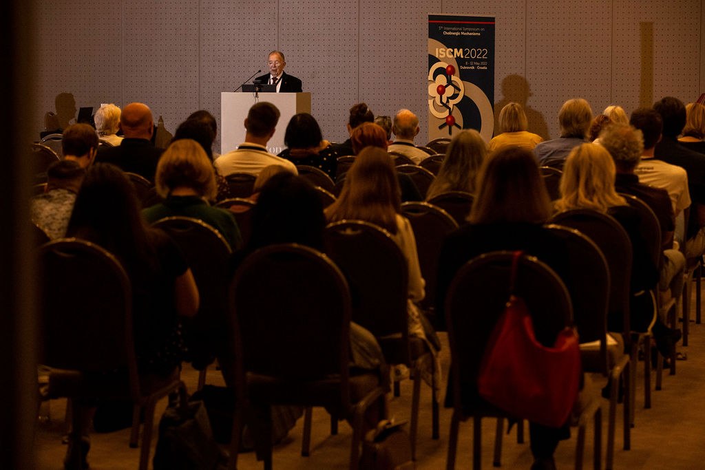
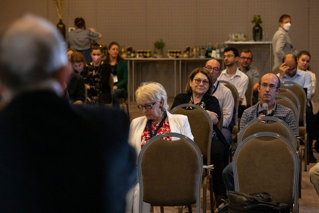
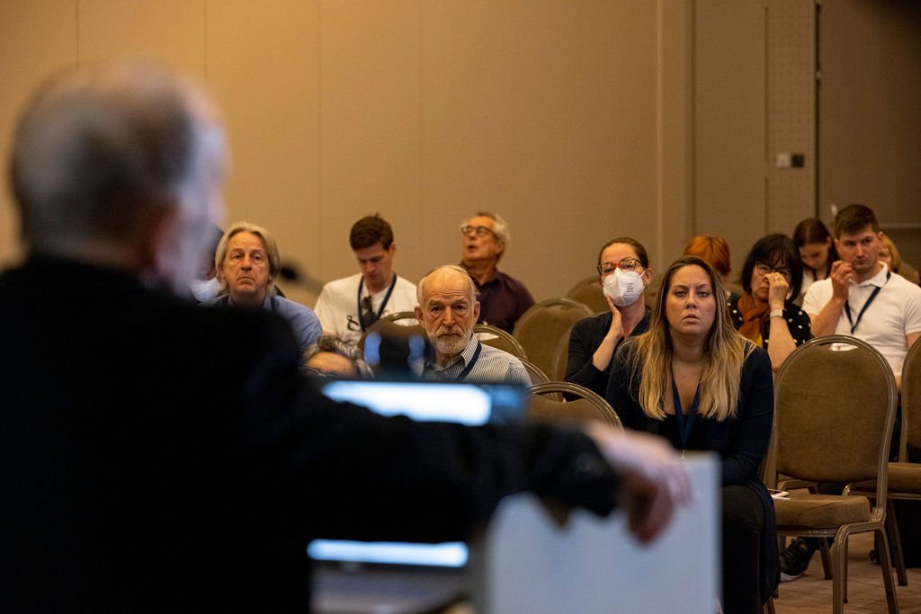
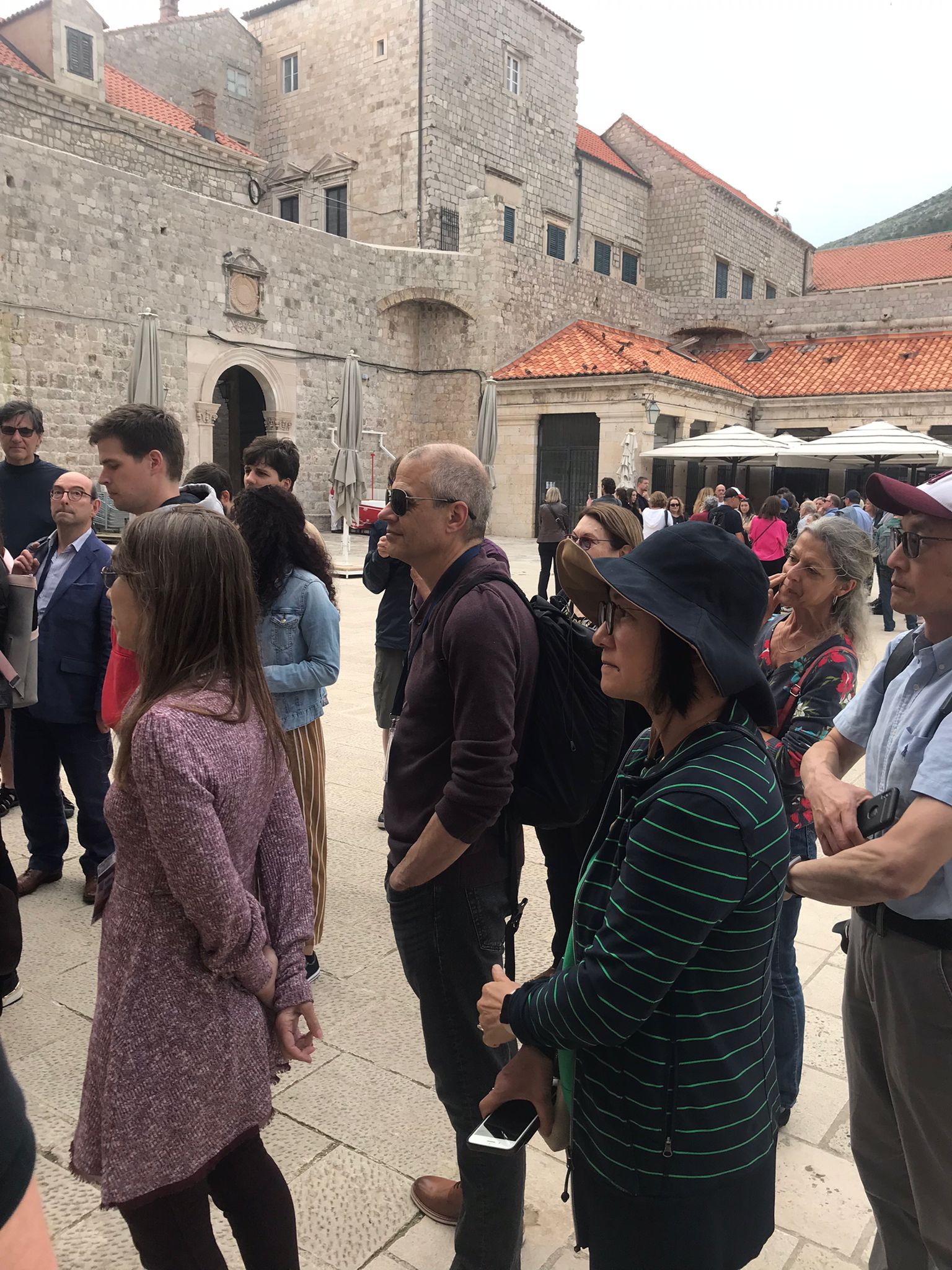
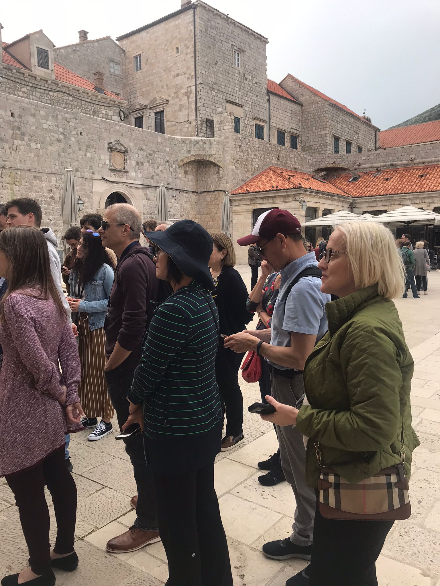
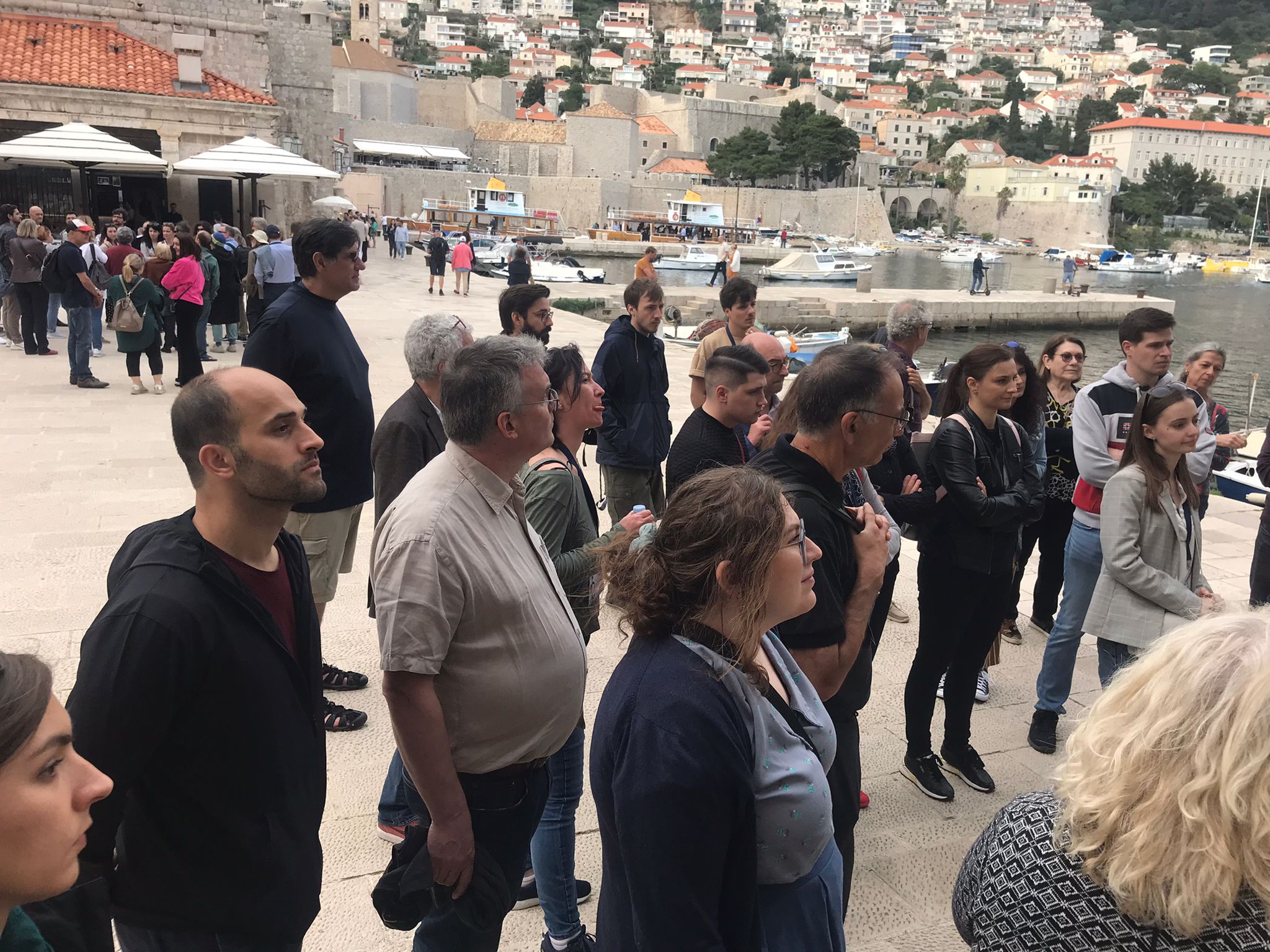
Participants
X


Proceedings:
The 17th International Symposium on Cholinergic Mechanisms