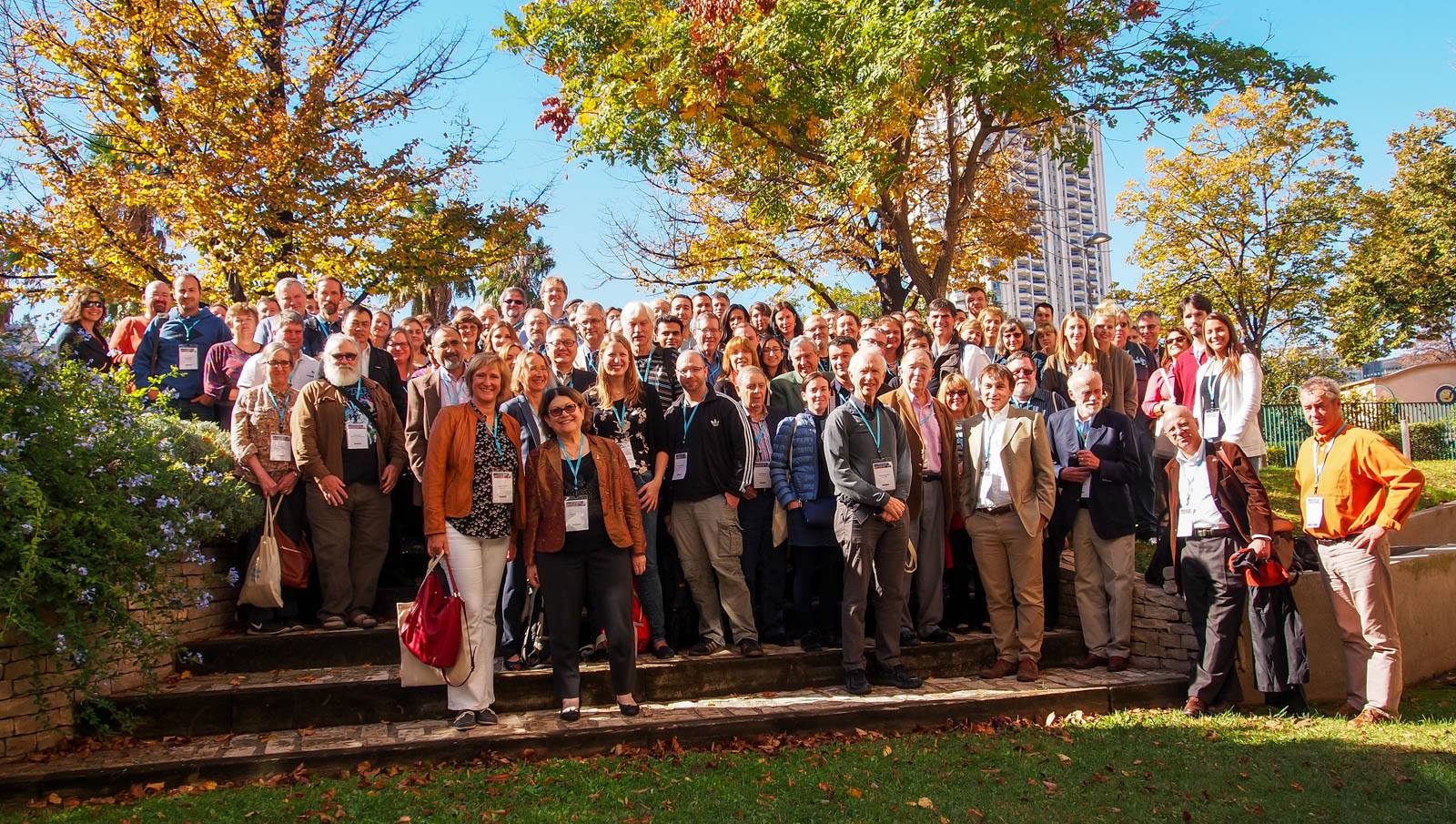
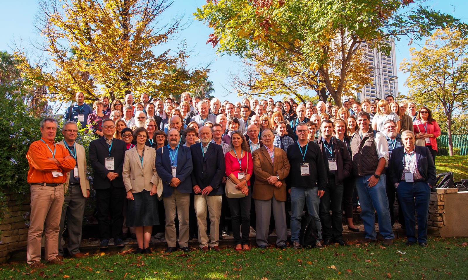
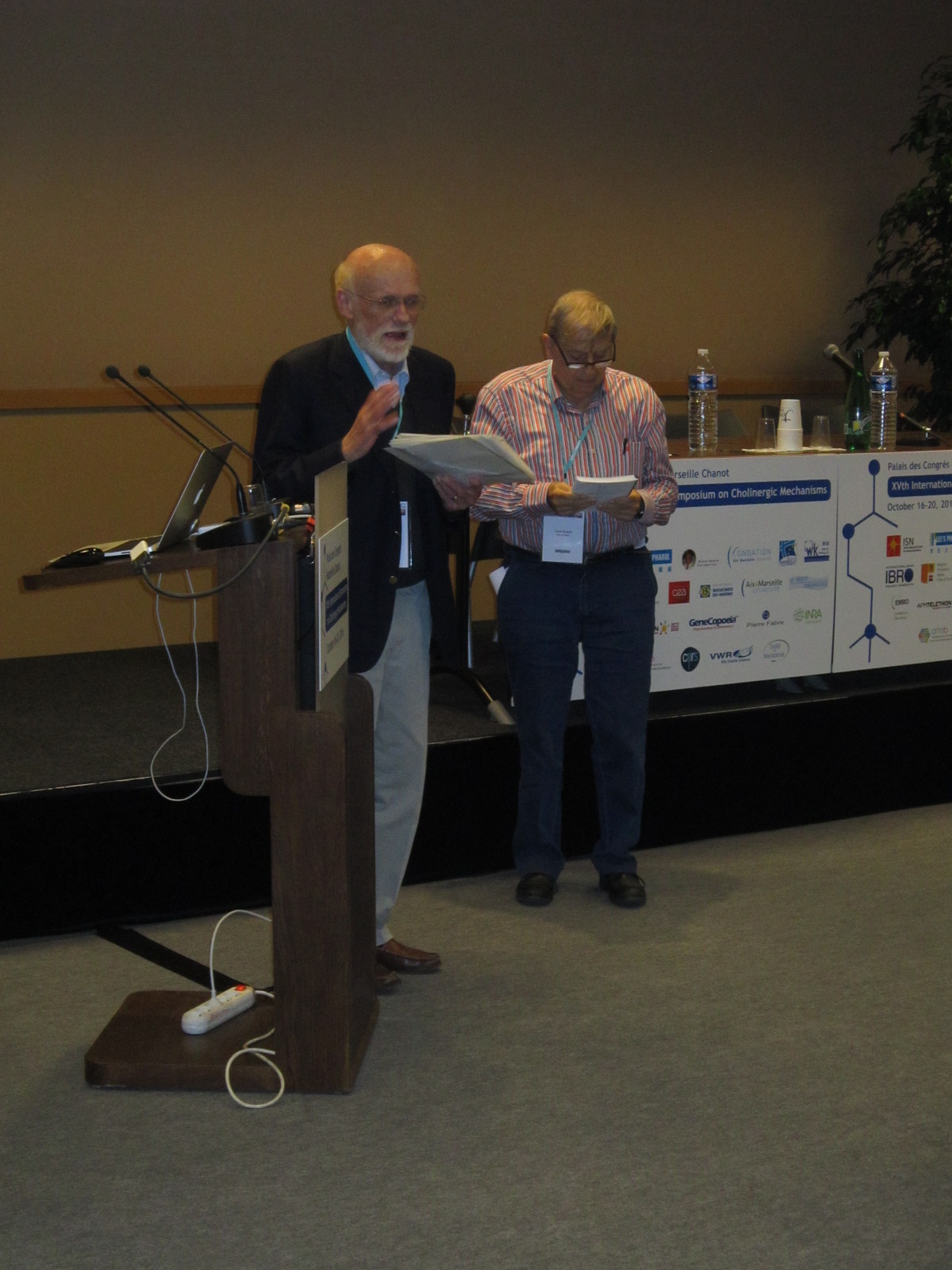
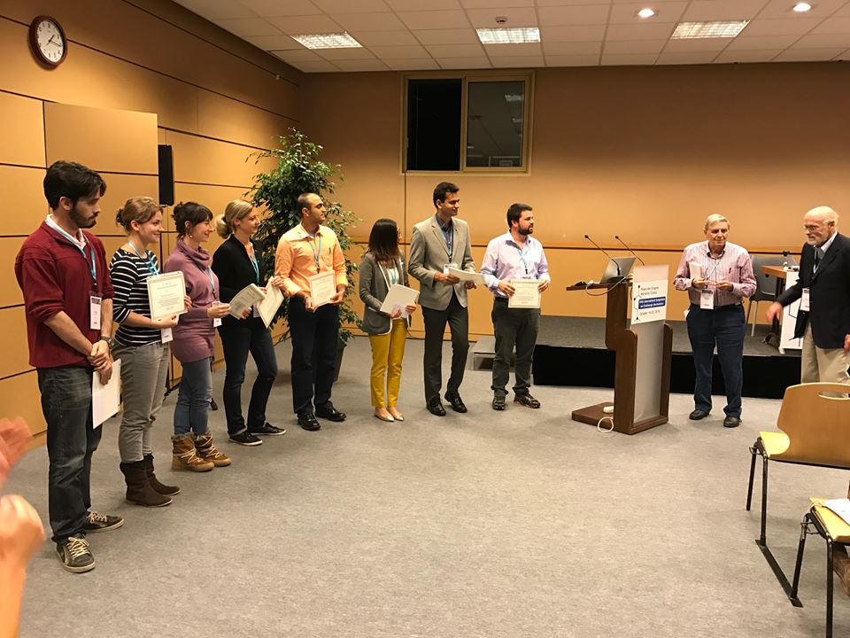
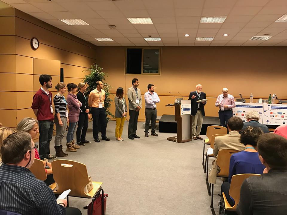
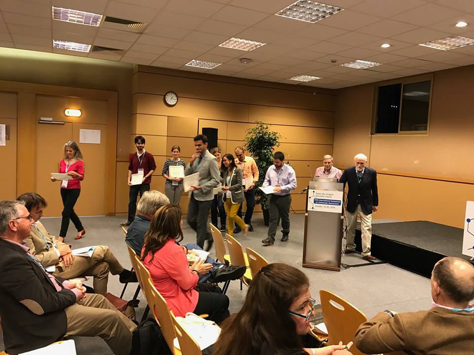
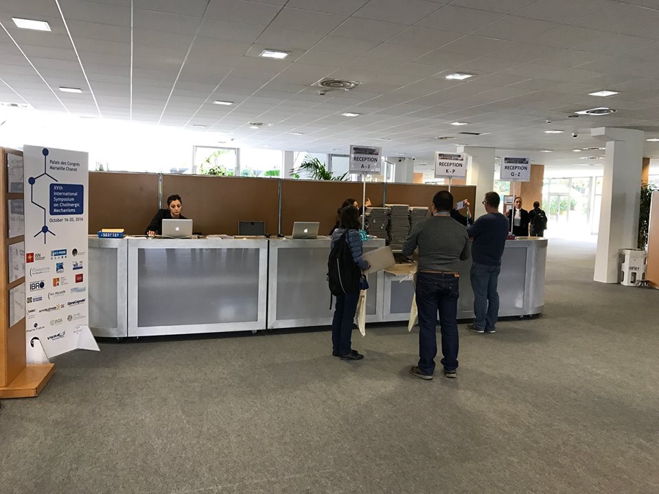
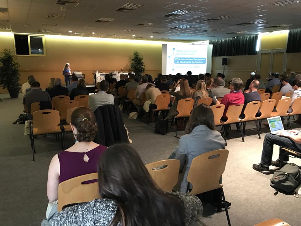
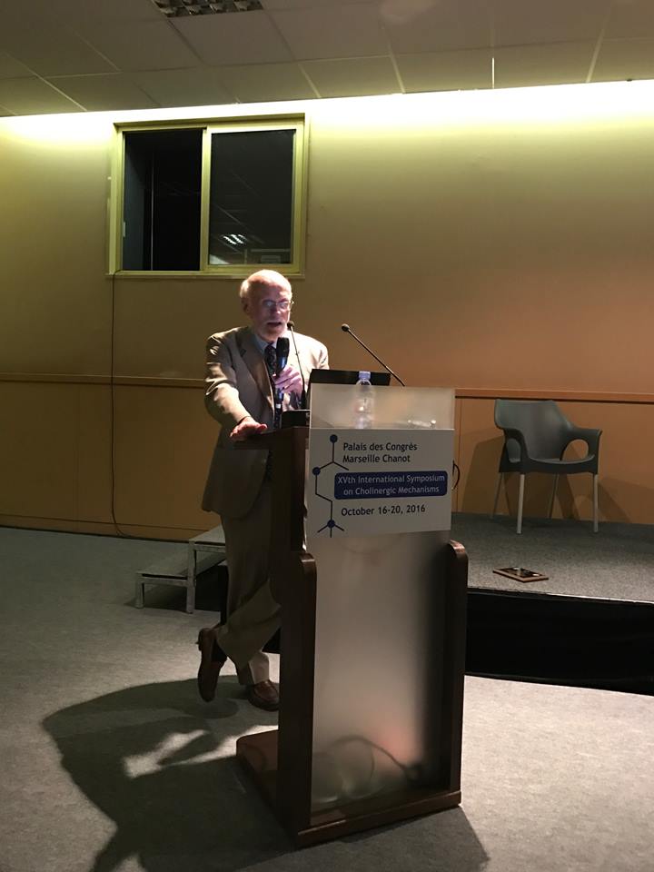
The 15th International Symposium on Cholinergic Mechanisms
2016 Marseille, France
The 15th International Symposium on Cholinergic Mechanisms
Proceedings:
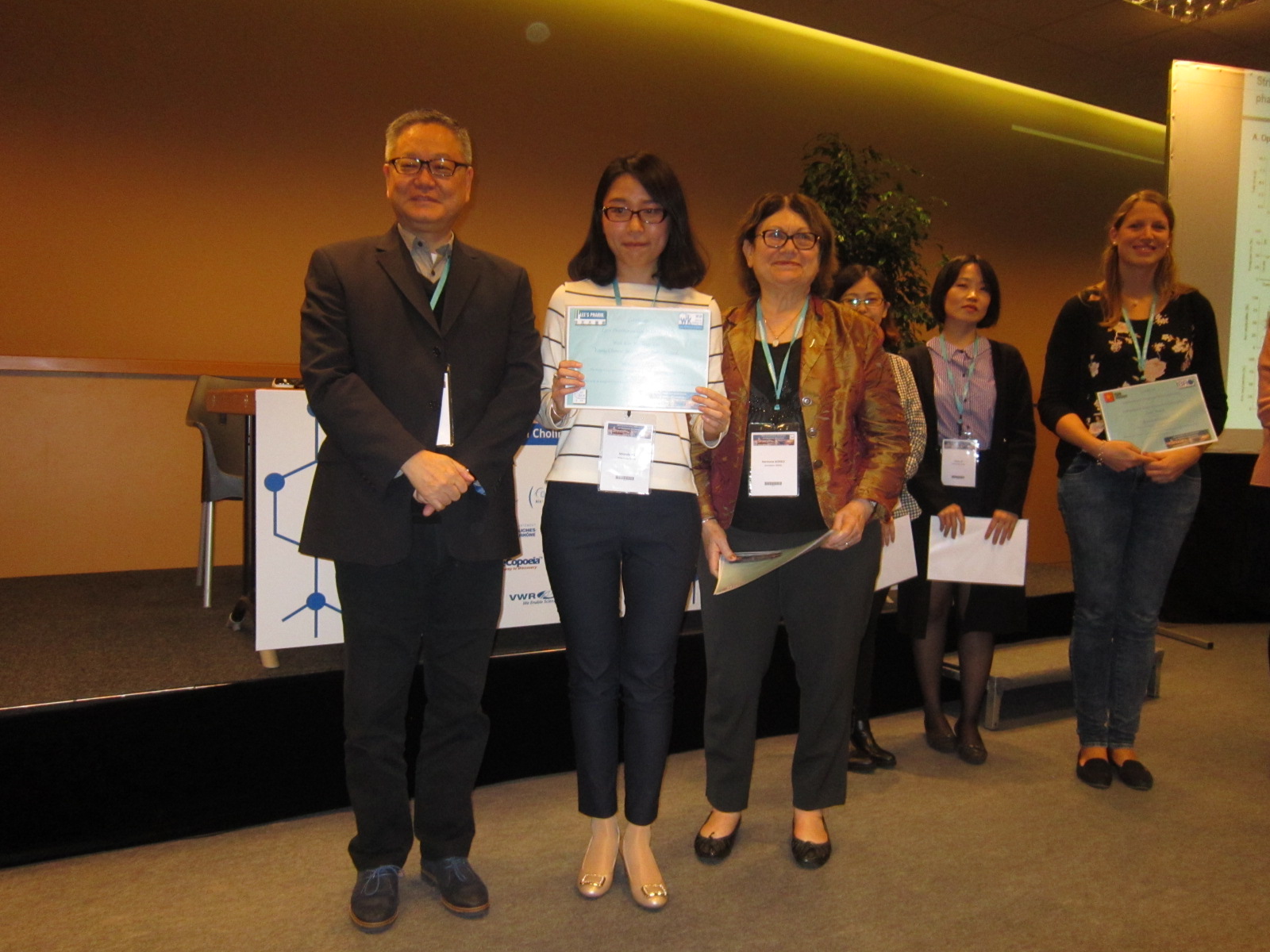
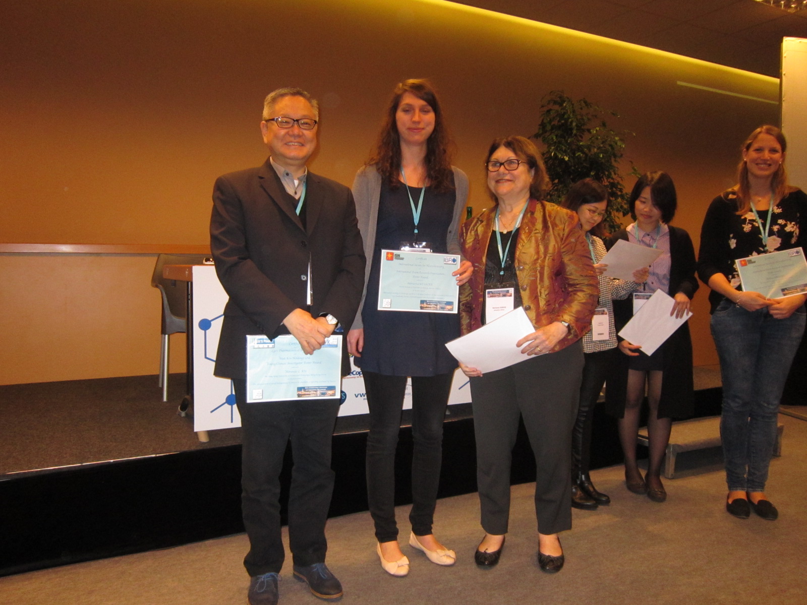
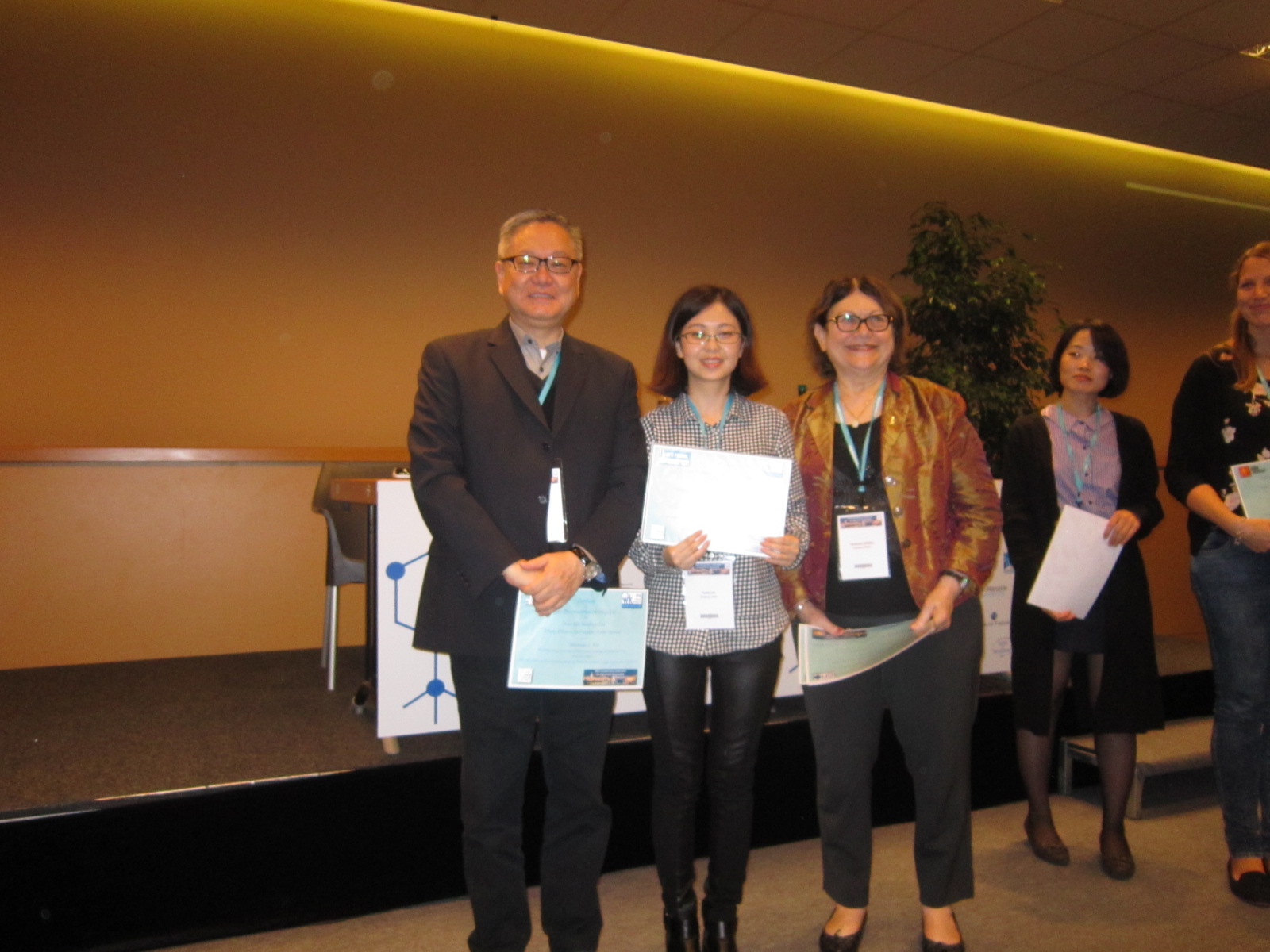
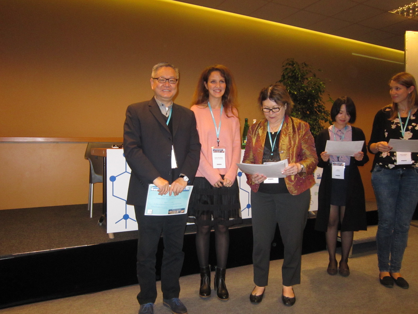
The 15th International Symposium on Cholinergic Mechanisms
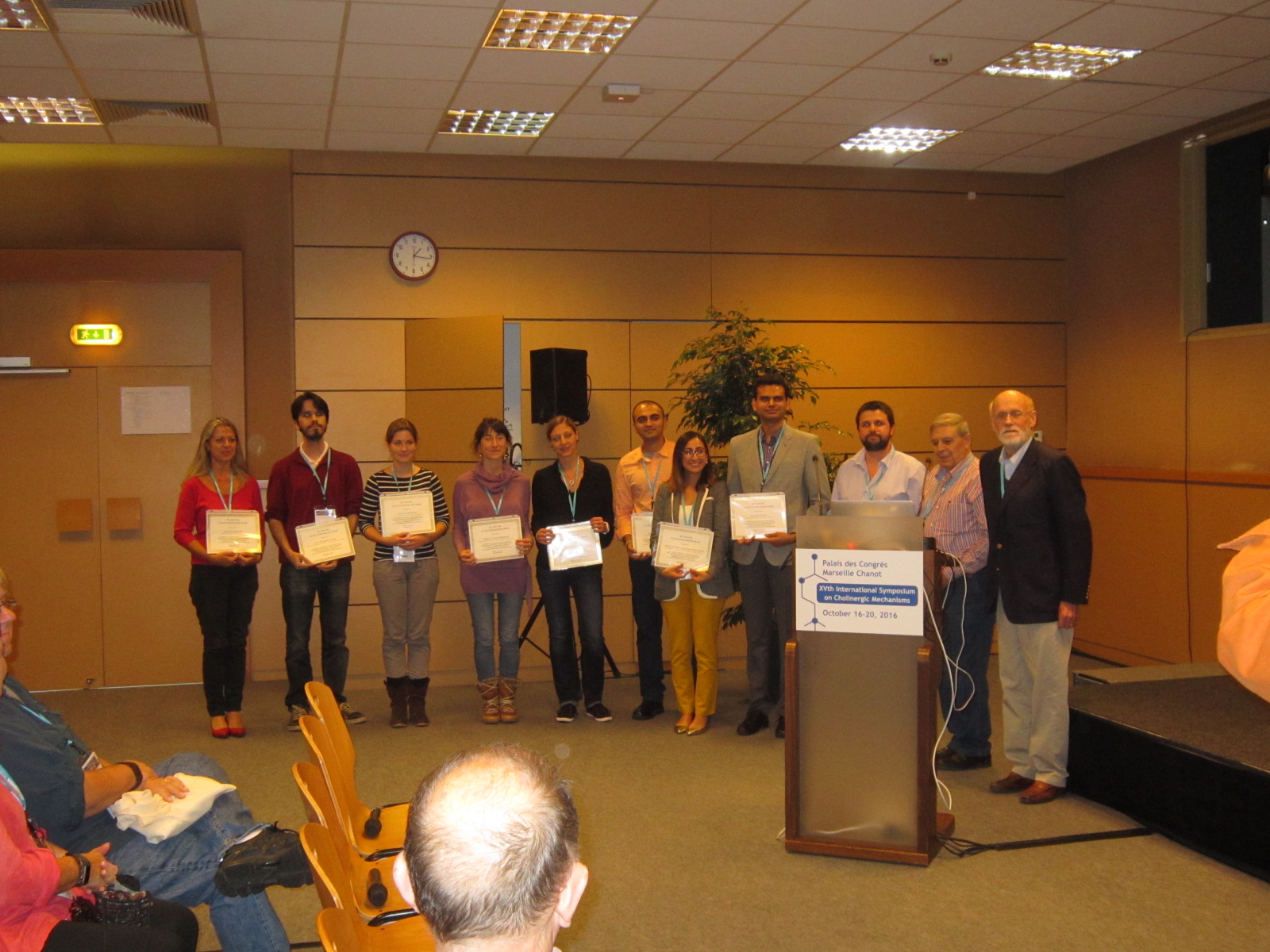
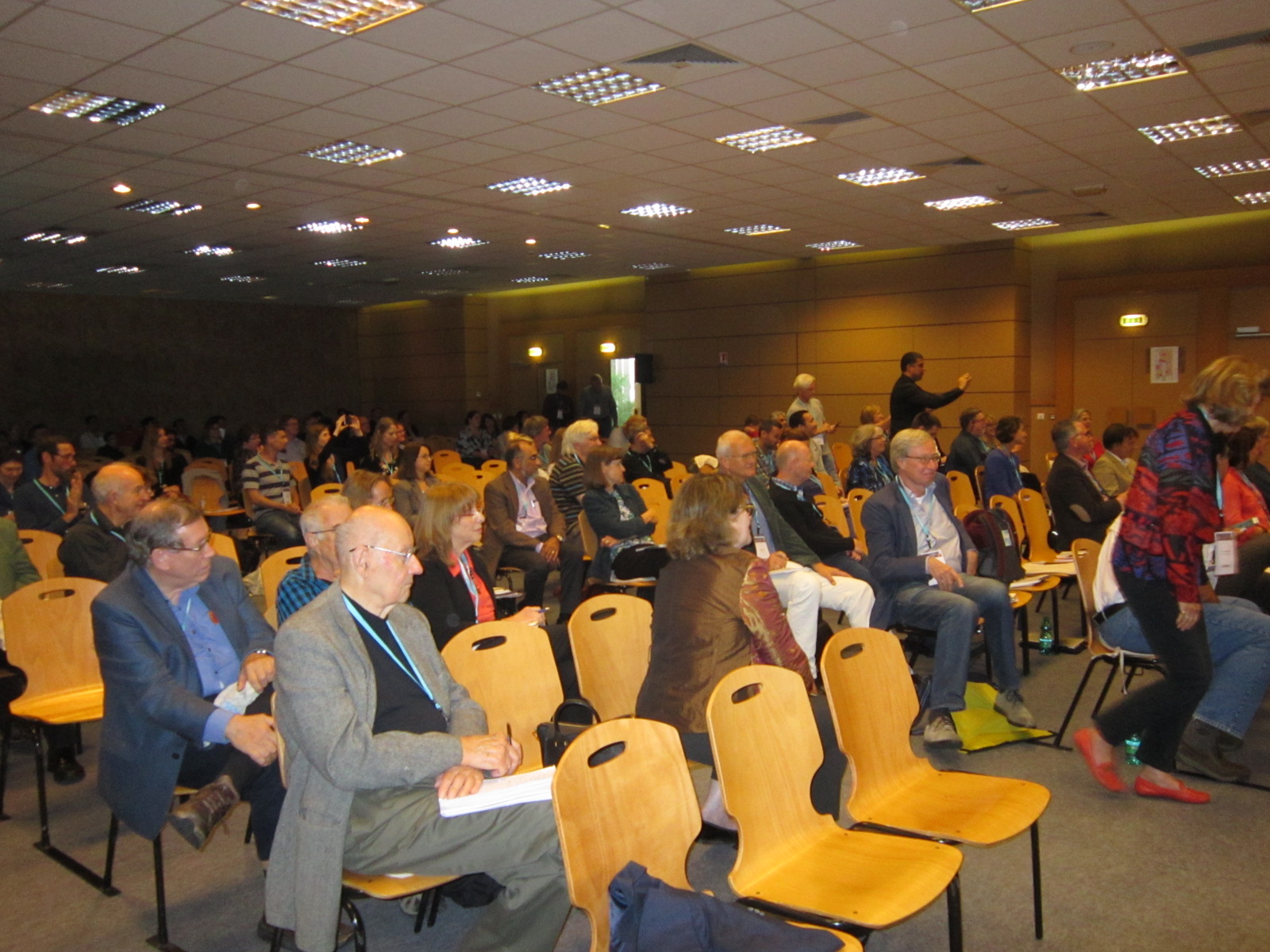
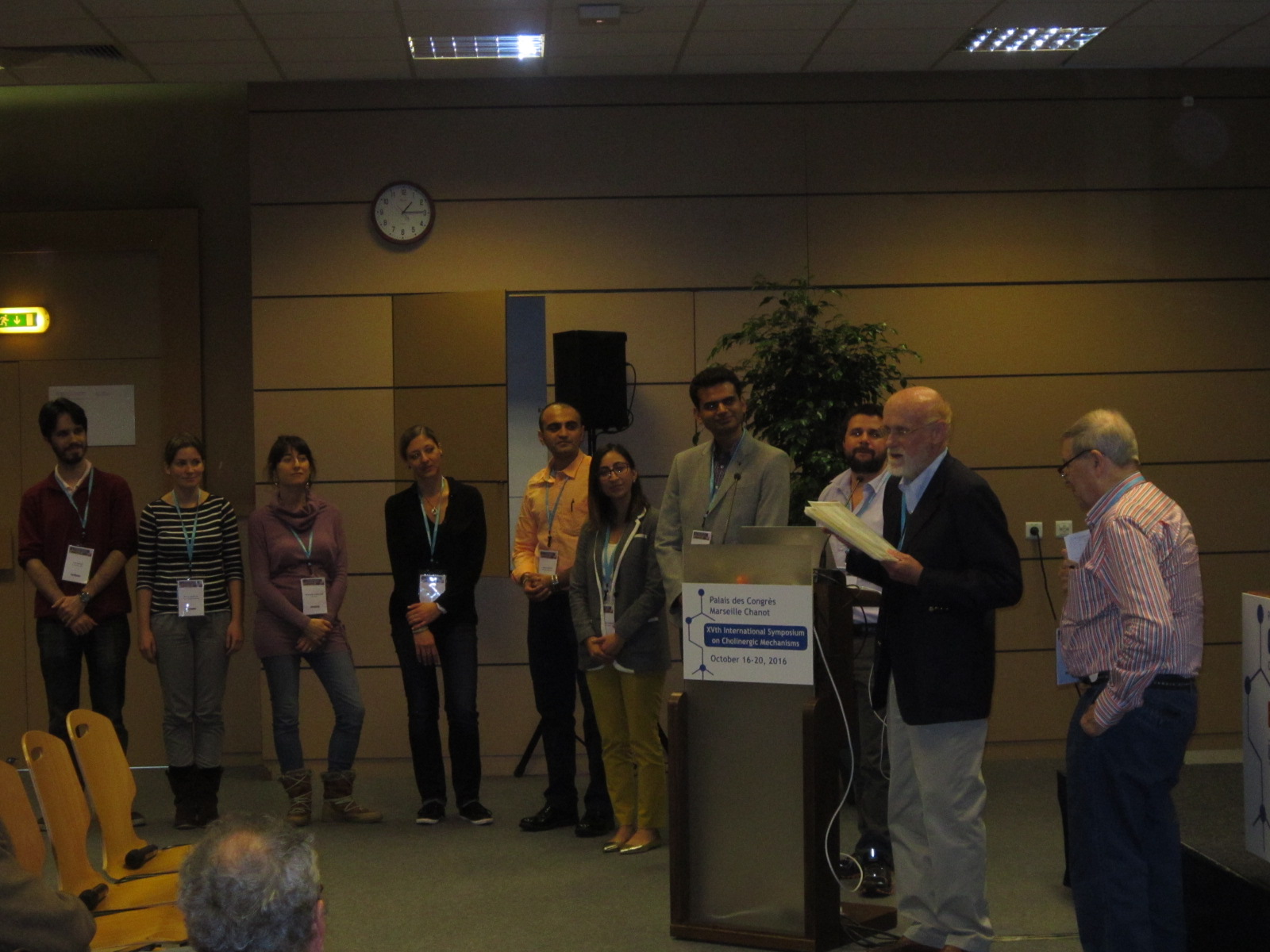
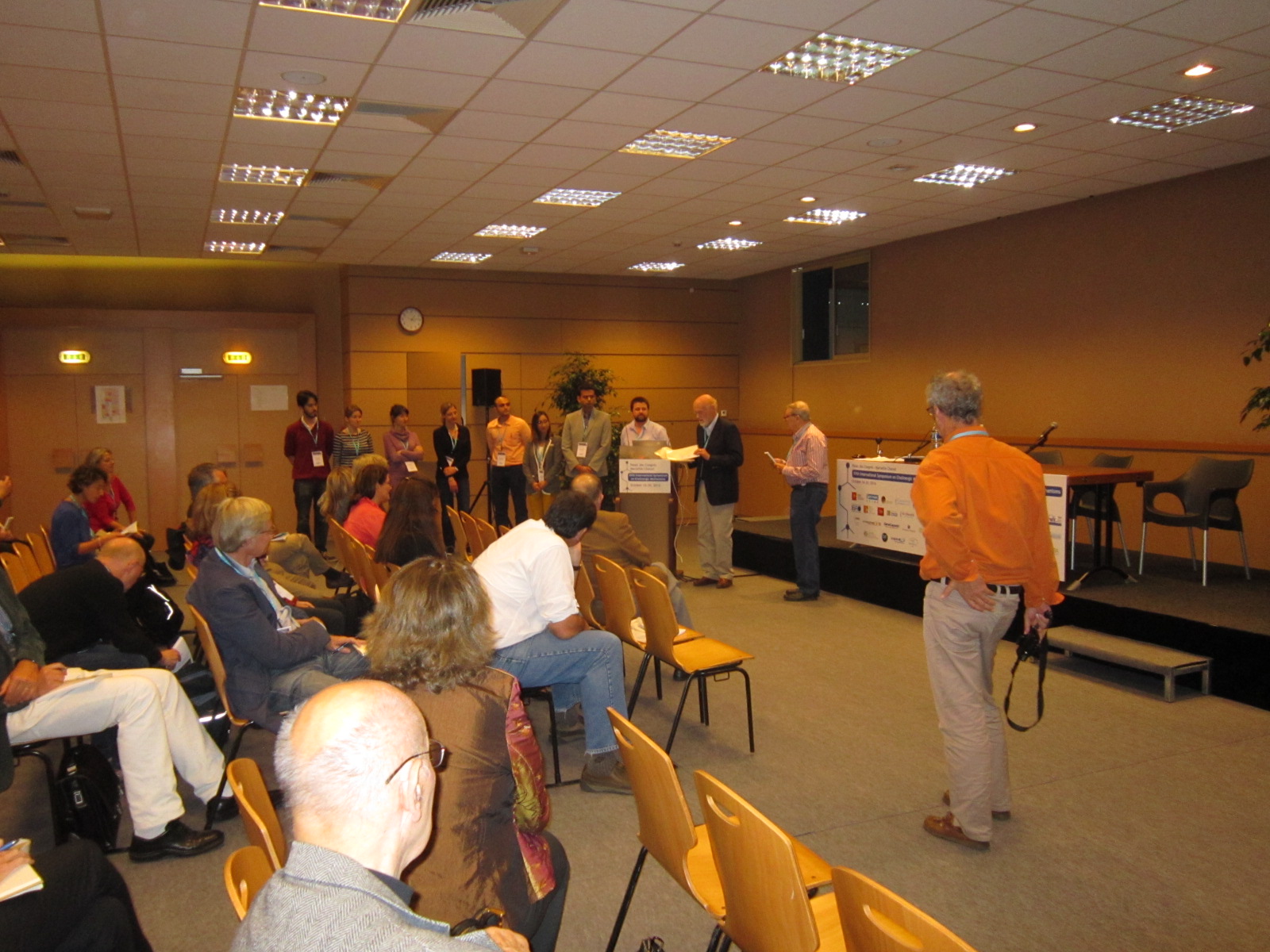
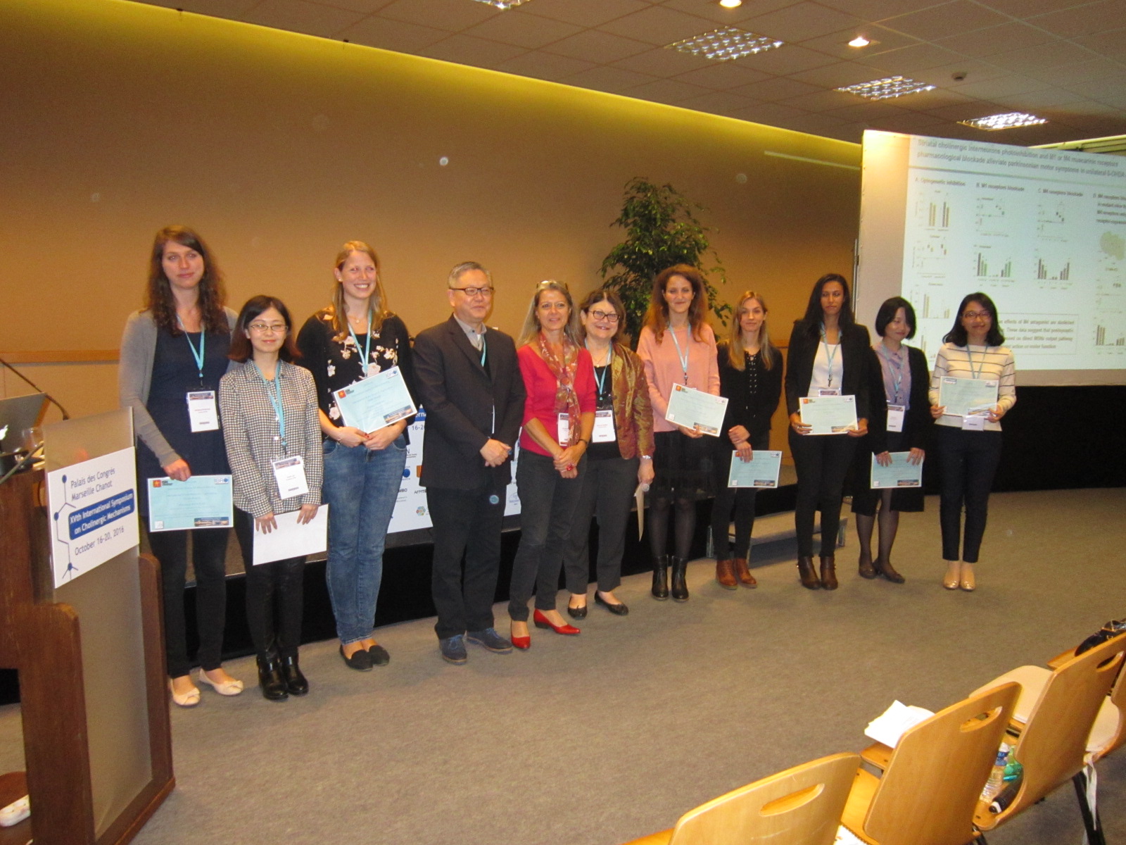
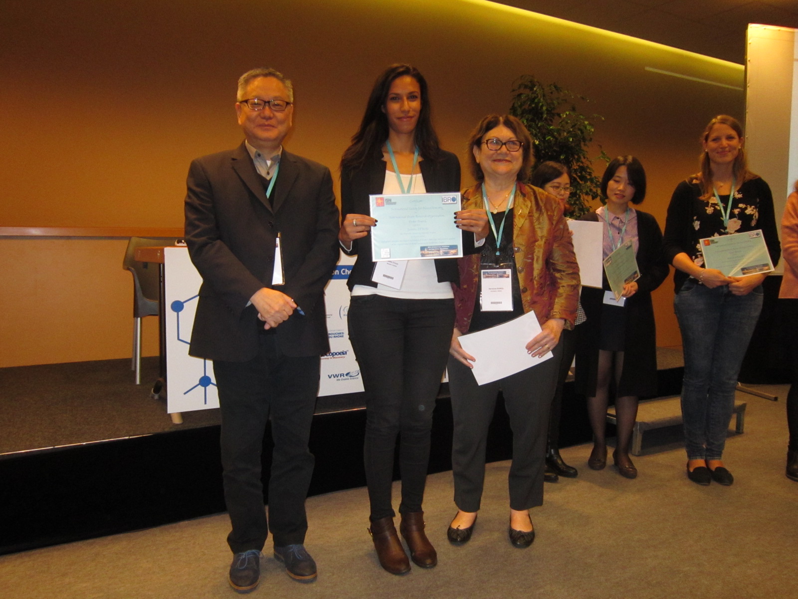
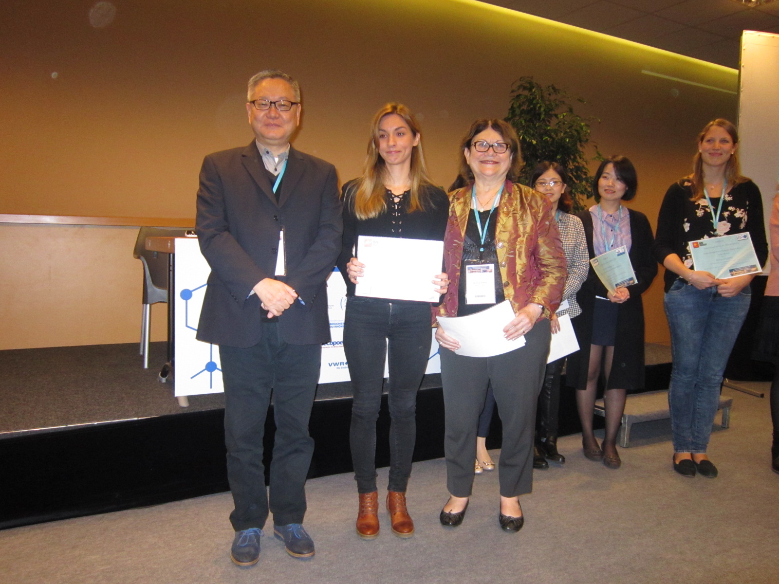
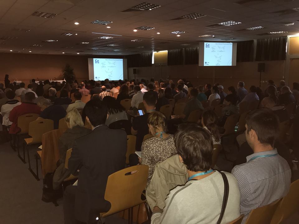
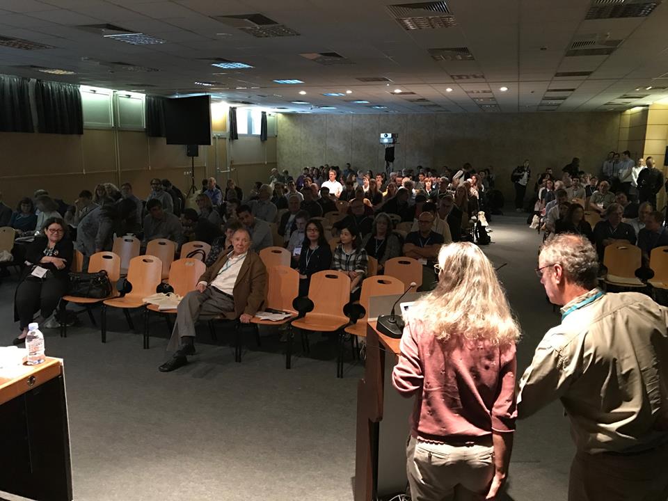
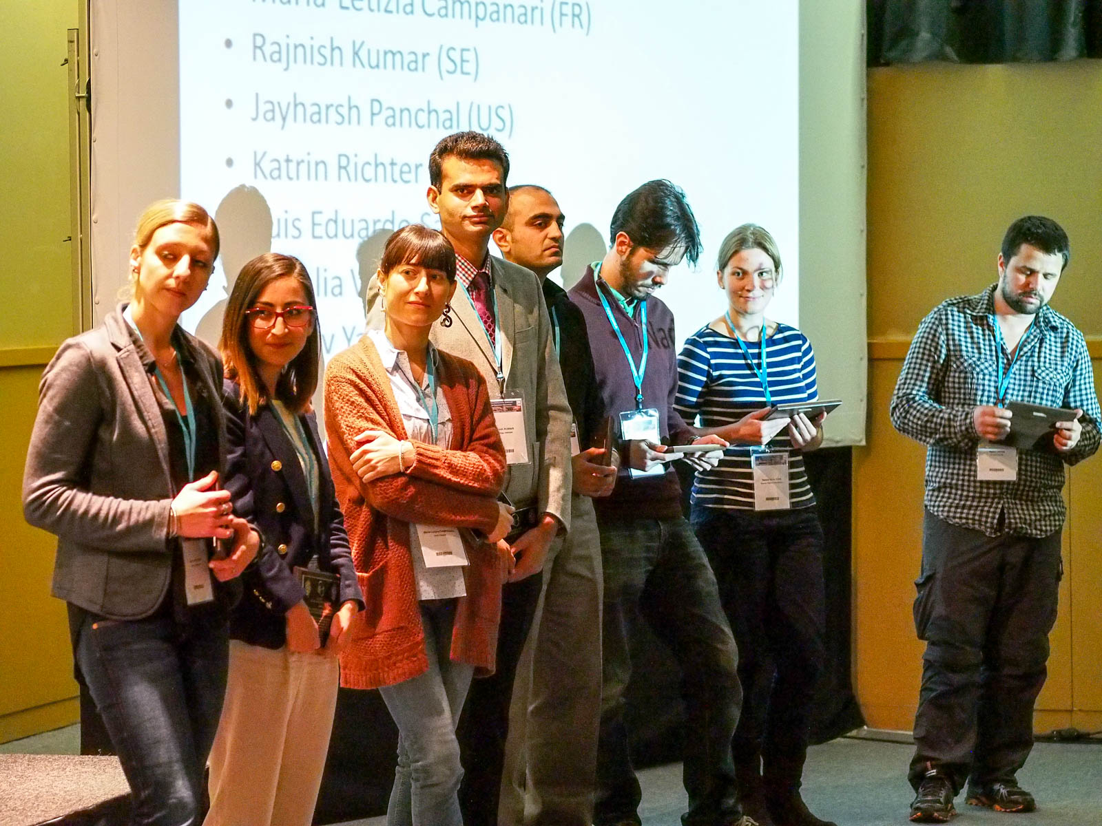
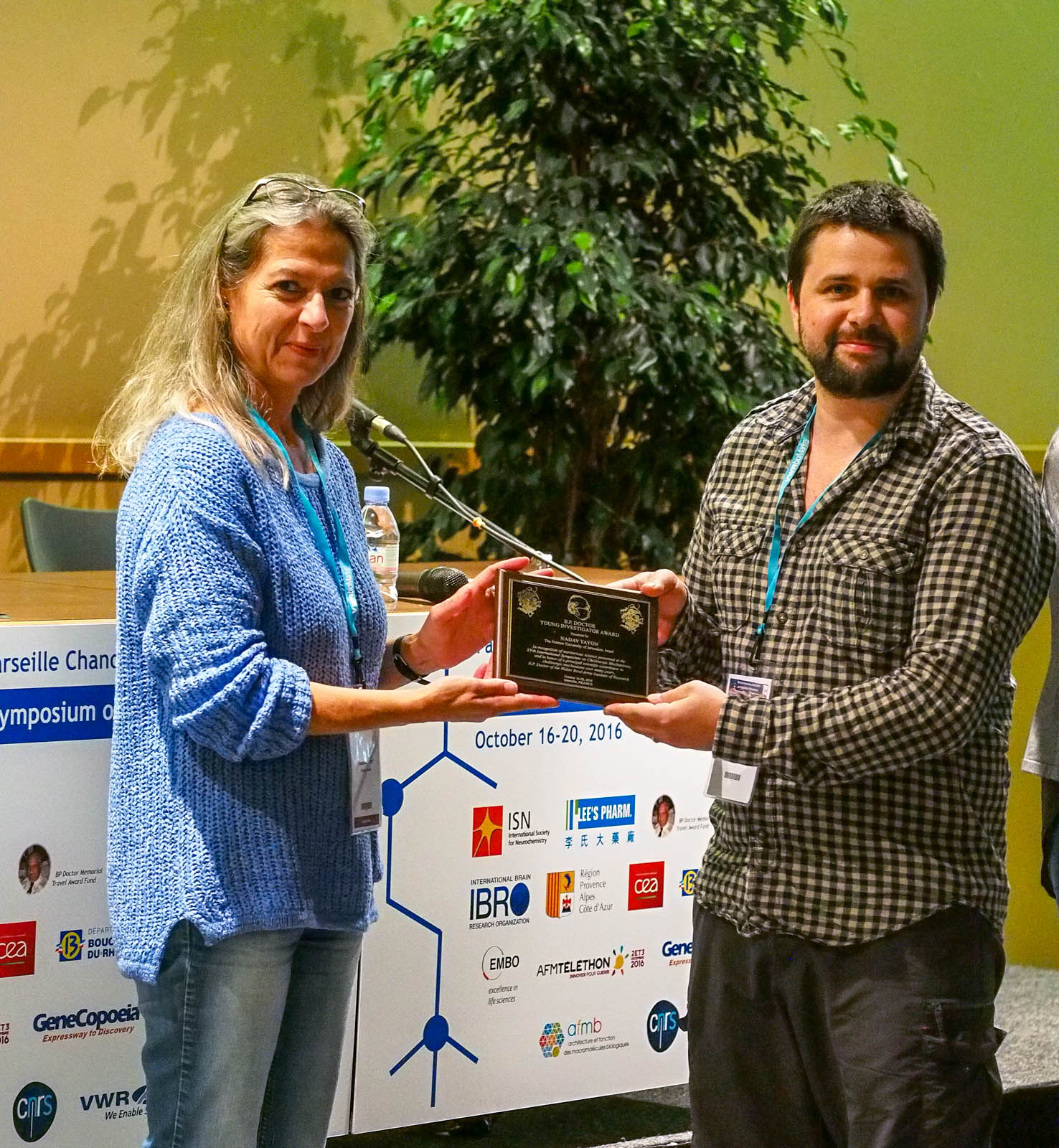
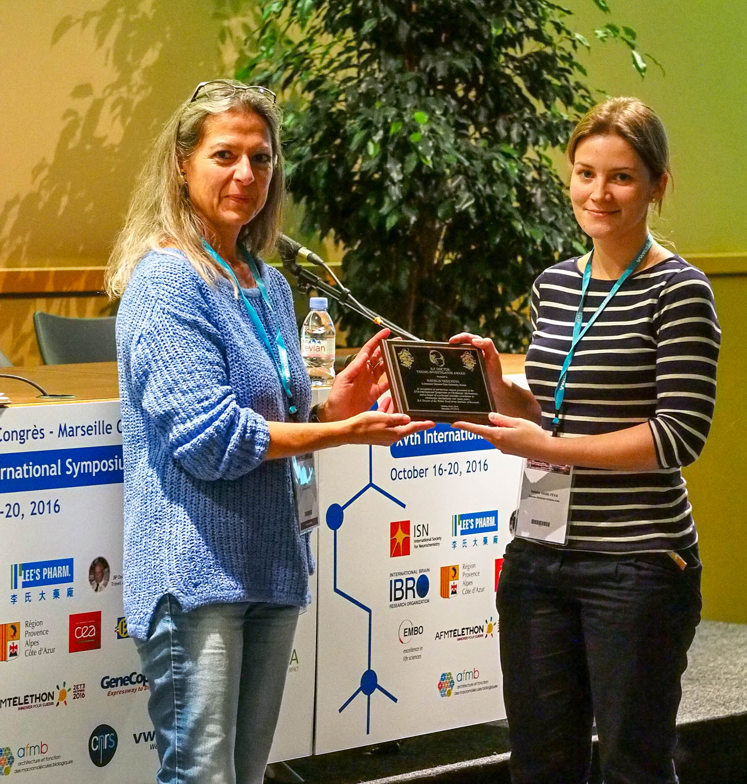
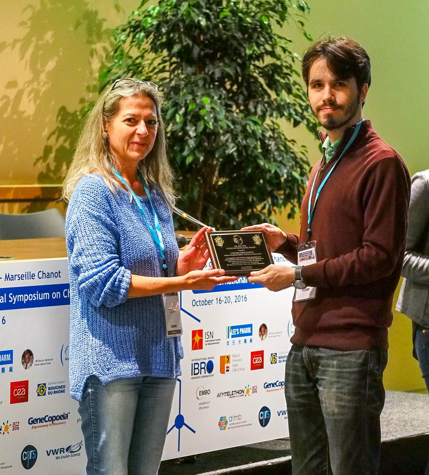
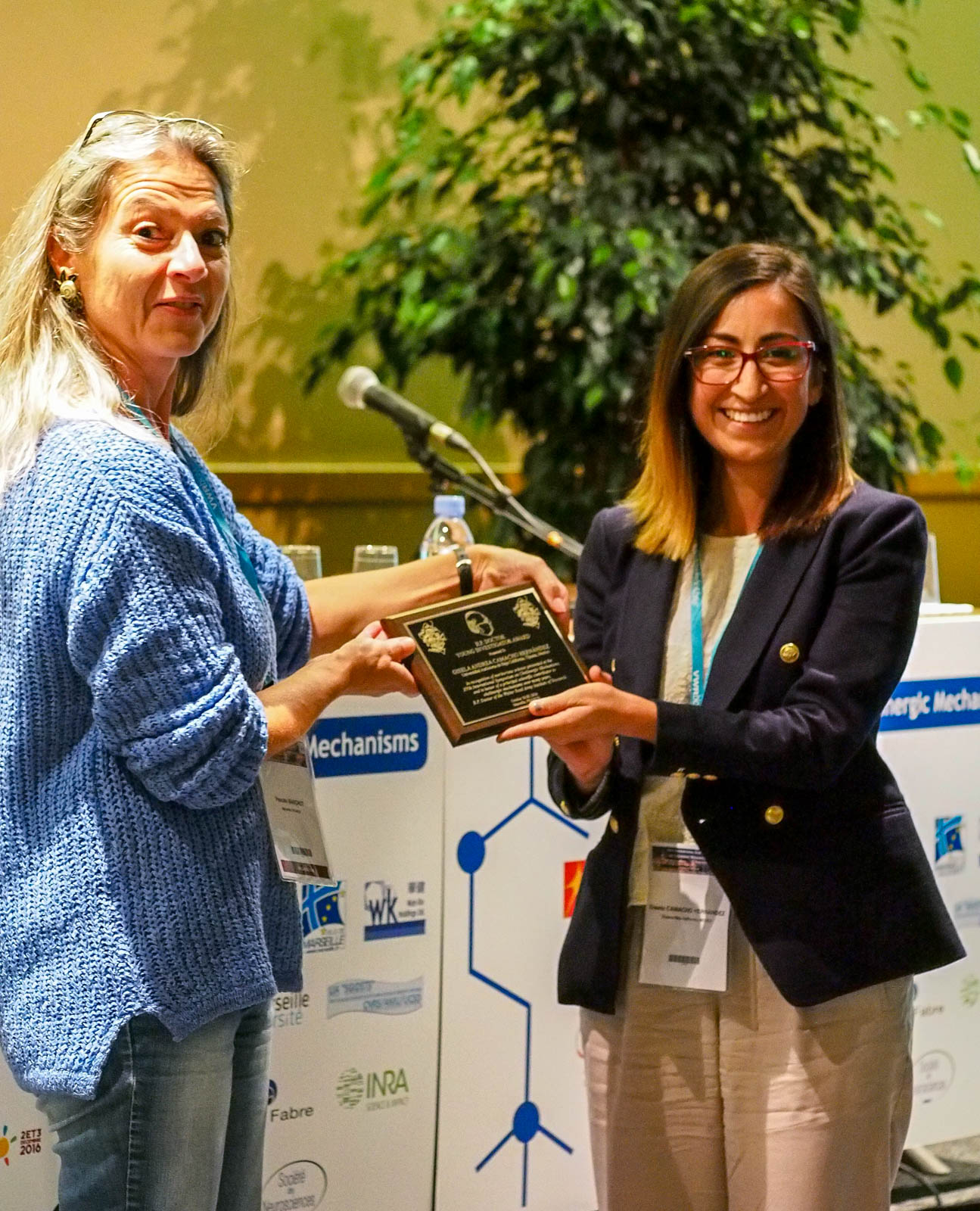
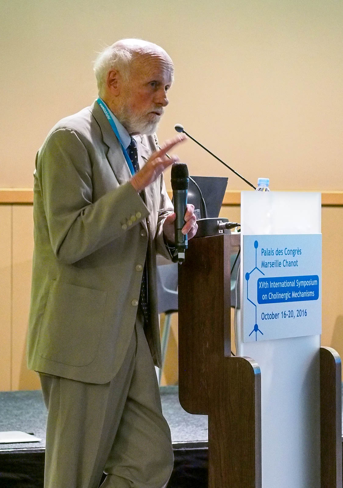
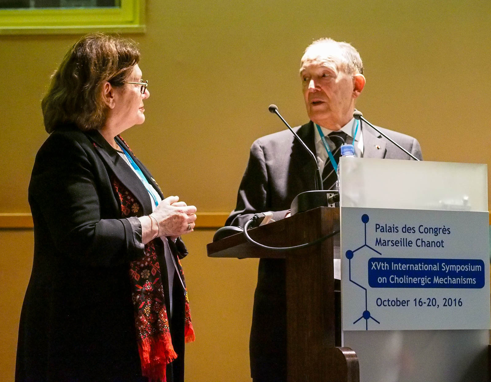
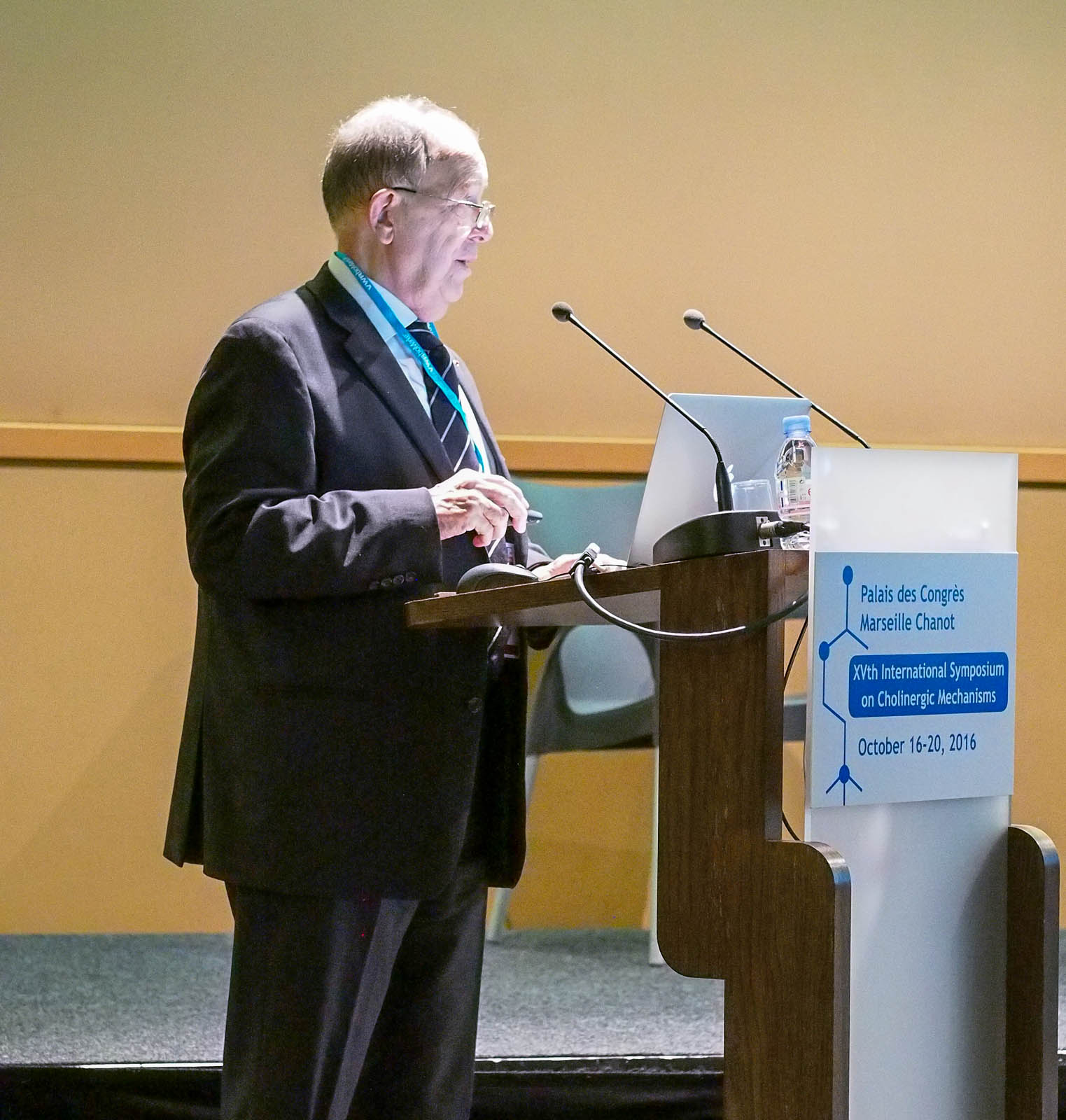
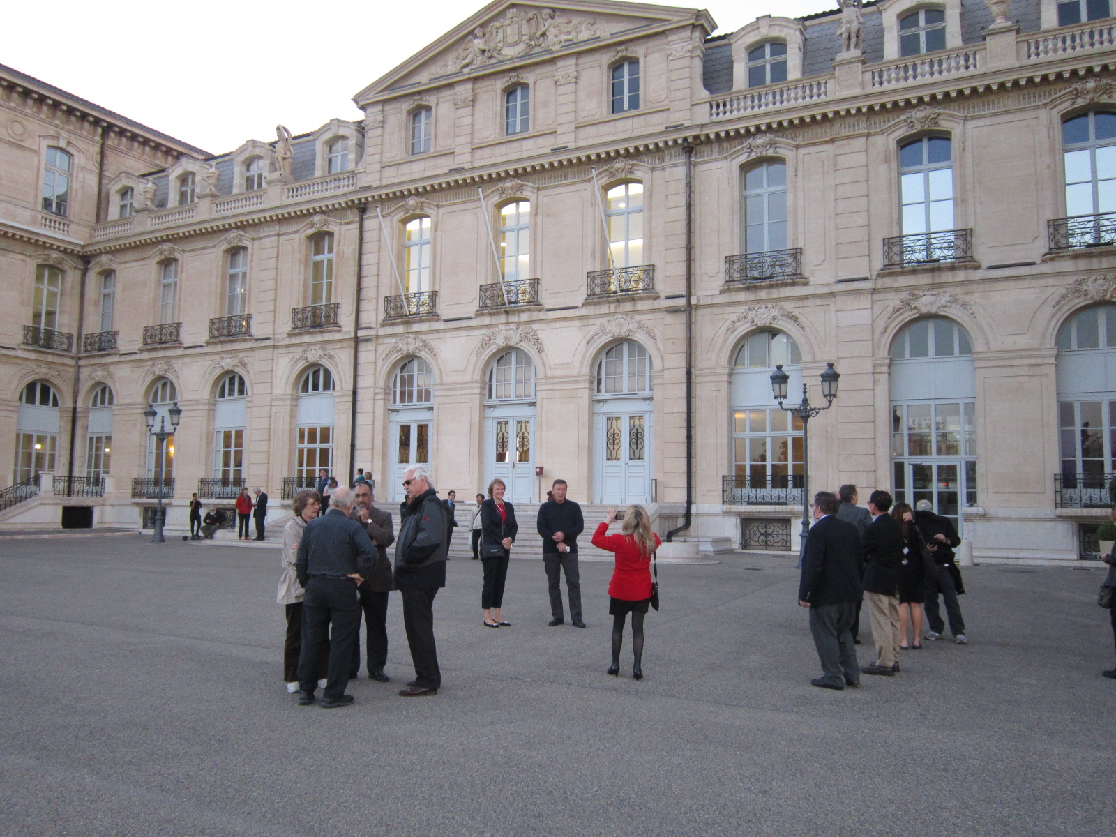
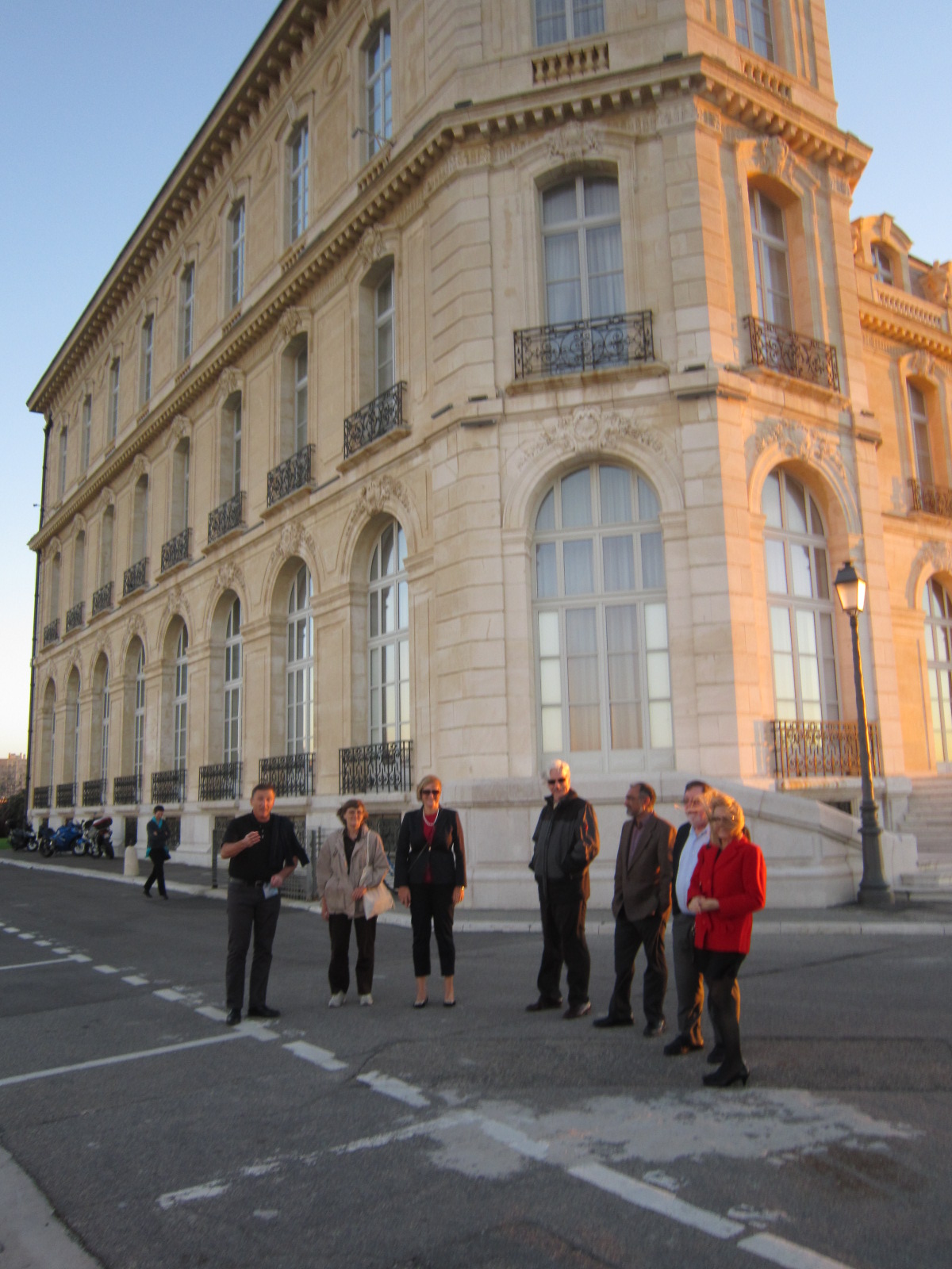
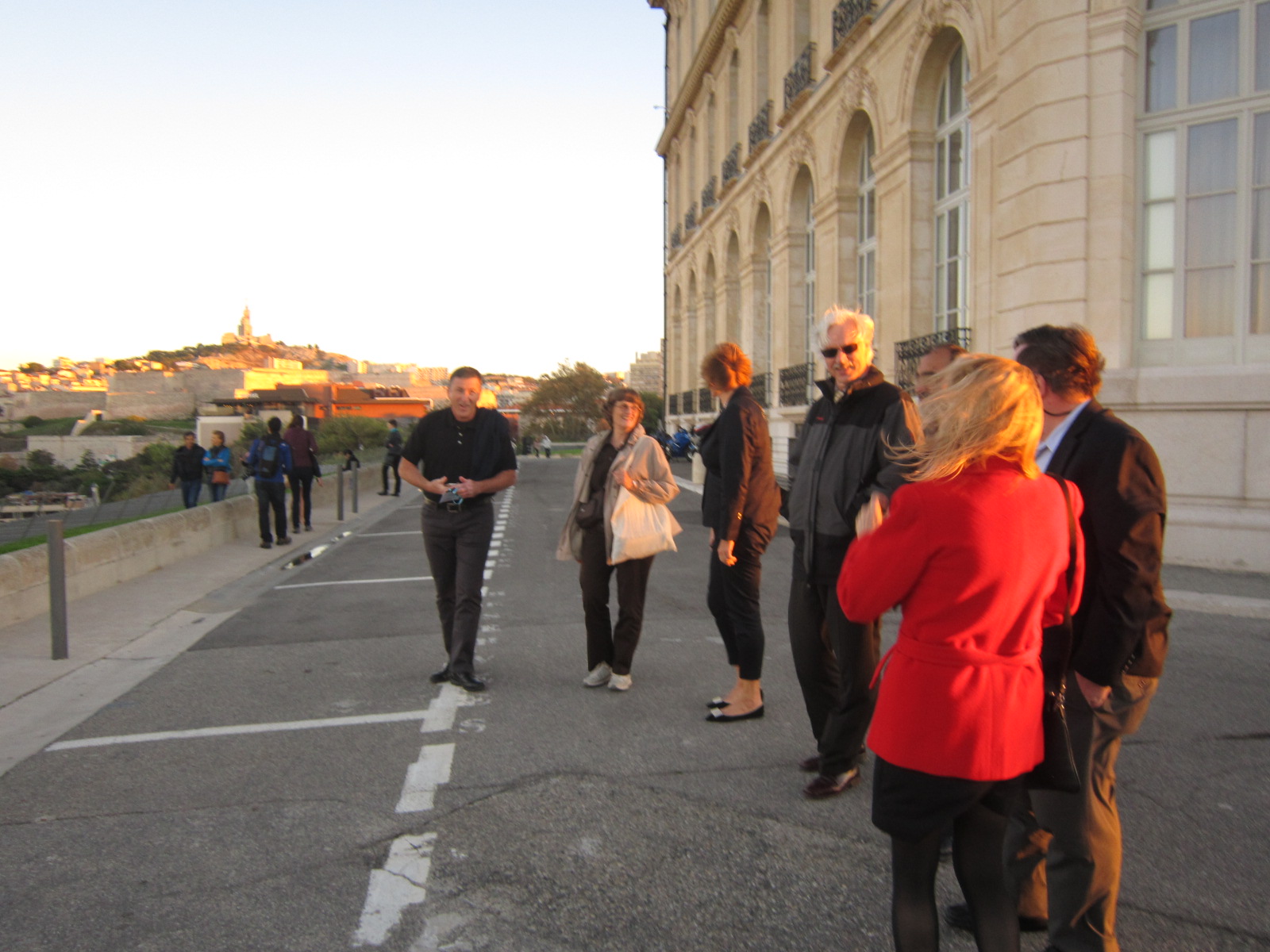
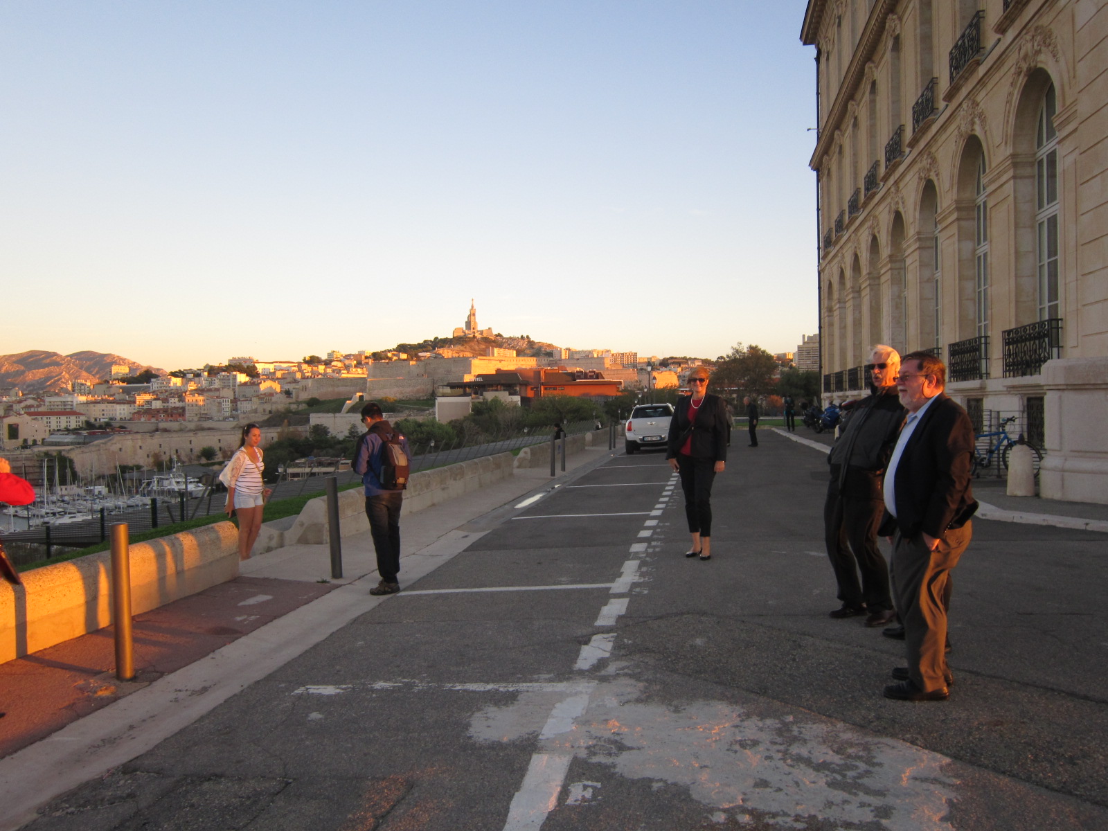
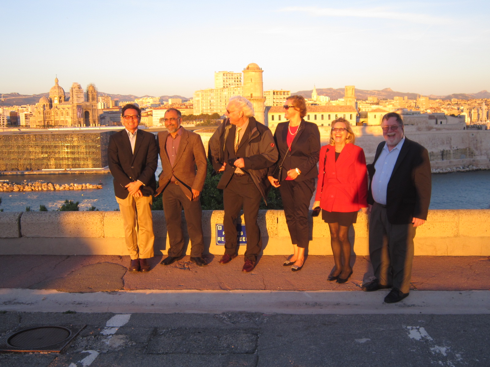
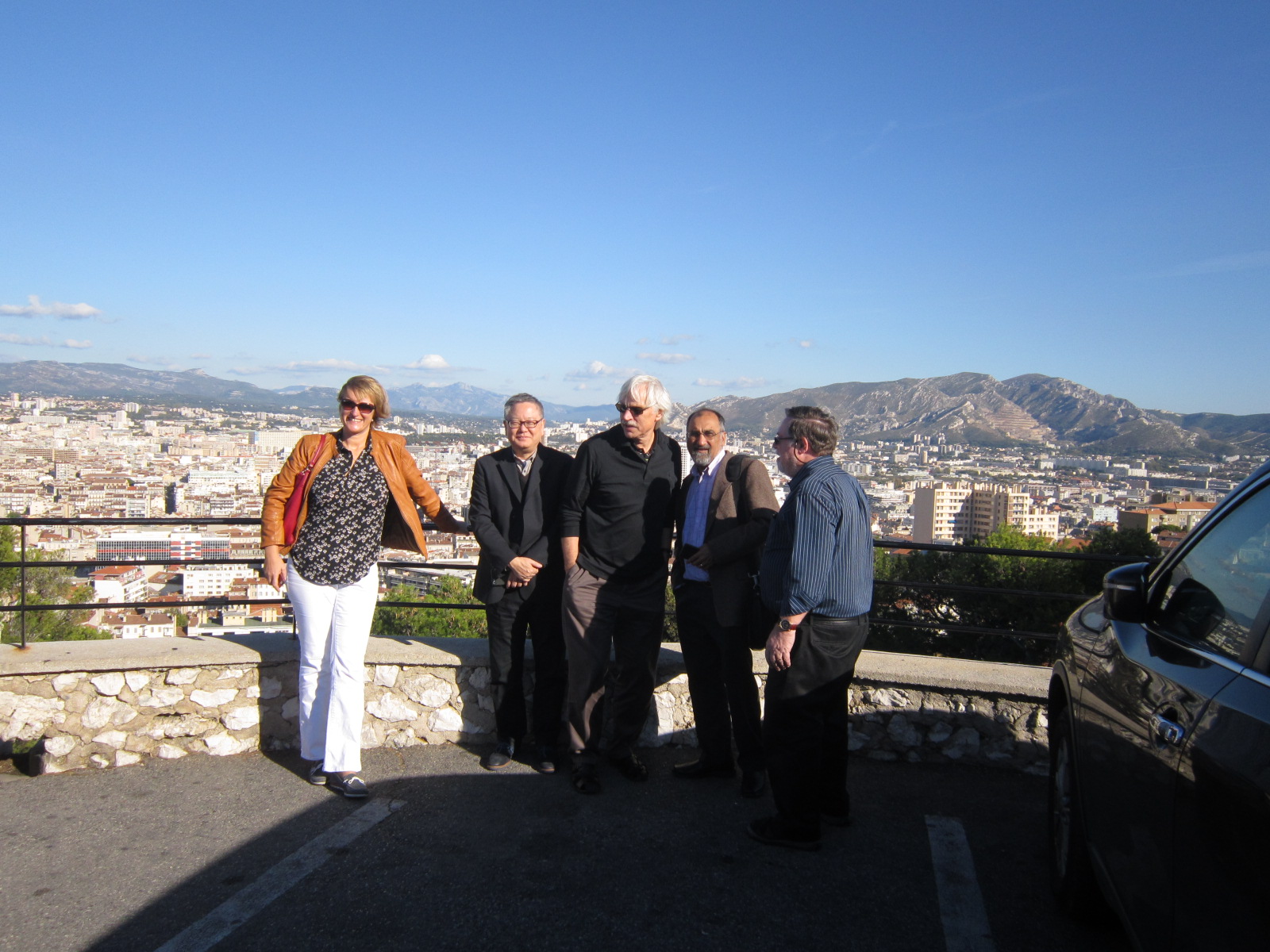
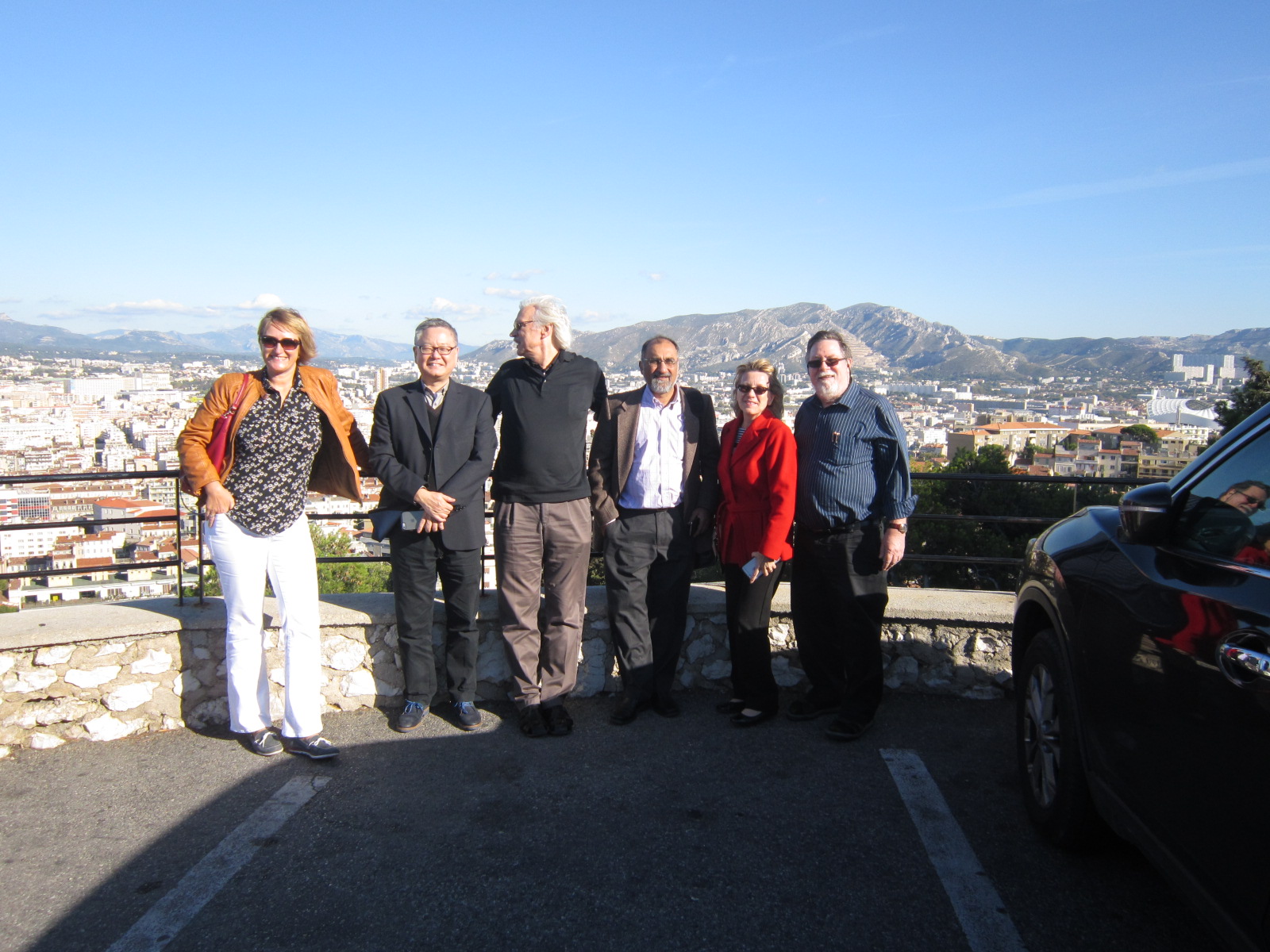
Participants
Q
U
Y